MRI · Spine
What a cervical spine MRI shows, and what the report means
Read Your Scan Editorial Team·Last updated
Written from published radiology references and reviewed for plain language. Not written or signed by a physician, and not a diagnosis. How we write these pages →
The short answer
A cervical spine MRI shows the seven neck vertebrae, the discs between them, the spinal cord running through the canal, and the nerve roots exiting at each level. Unlike the lower back, the cord itself is inside this region, so reports comment on it directly.
A cervical spine MRI makes detailed images of your neck — the bones, discs, spinal cord, and nerves. This page explains, in plain words, what it looks at and what common report terms mean.
What this scan shows
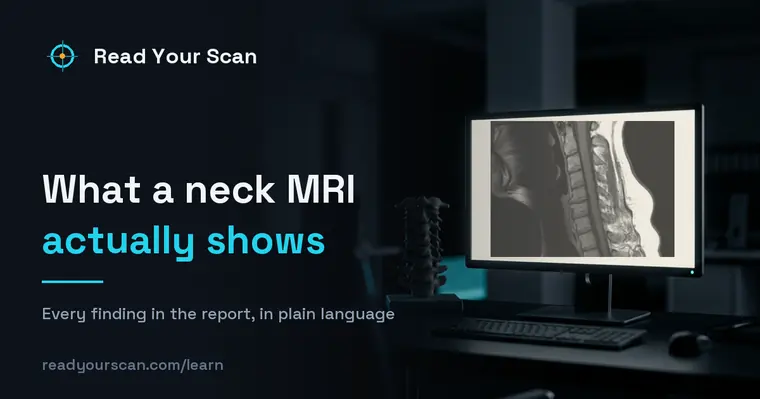
A cervical (neck) MRI shows the seven neck vertebrae, the discs between them, the spinal cord as it passes through, and the nerves that travel to the shoulders and arms. Doctors order it for neck pain, arm numbness or tingling, weakness, or after an injury. As with the low back, many findings it describes are common age-related changes seen in people without symptoms, so the report is read together with how you feel.

Common findings, in plain words
Disc bulge or herniation
Disc material extending beyond its normal edge. These are common in the neck and often cause no symptoms. Your doctor checks whether the location fits your arm or neck symptoms.
Cervical spondylosis
A general term for age-related wear of the neck’s discs and joints. It is very common and often described simply as part of the picture.
Foraminal narrowing
Narrowing of the small openings where nerves leave the spine. It can be mild to severe, and your doctor relates it to any arm symptoms.
Spinal cord signal change (myelomalacia)
An area where the spinal cord looks slightly different on the images. It is described carefully so your doctor can consider its meaning alongside your exam.
Osteophytes (bone spurs)
Small bony growths that form with wear over time. They are common and often cause no trouble on their own.
Straightening of the normal curve
The usual gentle neck curve appearing flatter, sometimes linked to muscle tightness. It is a frequent, often minor observation.
Uncovertebral arthrosis
Wear in the small joints along the sides of the neck vertebrae. It is very common with age and matters mainly when the overgrown bone narrows the channel a nerve root leaves through.
Terms you might see
- Cervical
- Referring to the neck region of the spine, with vertebrae labeled C1 through C7.
- Spinal cord
- The bundle of nerves running through the spine that carries signals between the brain and body.
- Osteophyte
- A bone spur — a small extra bit of bone that grows with age-related wear.
- Foramen
- The opening on each side of the spine where a nerve exits toward the arm.
- Radiculopathy
- A word describing symptoms — such as arm pain or tingling — that can occur when a nerve root is irritated.
Questions to ask your doctor
- 1.Do the MRI findings match where I feel neck or arm symptoms?
- 2.Are these changes typical for my age, or does anything stand out?
- 3.What non-surgical approaches, like physical therapy, might help?
- 4.Are there symptoms that would mean I should be seen more urgently?
- 5.Do I need any follow-up imaging, and when?
See these findings on your own scan
Upload your scan (de-identified in your browser) and your radiologist’s report, and every finding is mapped onto the images, explained in plain language and scored — free, no card needed. Informational only — not a diagnosis.
FAQ
- My neck MRI mentions spondylosis — what is that?
- Spondylosis is a general term for normal age-related wear of the discs and joints in the neck. It is extremely common and often causes no symptoms. Your doctor interprets whether it relates to how you feel.
- Does neck numbness in my arm always come from the MRI findings?
- Not necessarily. Arm symptoms can have several causes. The MRI is one piece of information your doctor combines with your exam and history to understand what is going on.
- Should I be worried about bone spurs?
- Bone spurs (osteophytes) form gradually with age and are very common. Many cause no problems at all. Your doctor considers them in the context of your overall picture.
Related
Go deeper
Sources
- Patient-facing procedure descriptions (MRI, CT, X-ray, ultrasound)RadiologyInfo.org — Radiological Society of North America & American College of Radiology
- MRI scan — overview, how it is performed and resultsNHS (United Kingdom)
- ACR Appropriateness Criteria and practice parameters for reportingAmerican College of Radiology
Read Your Scan is informational only — not a medical diagnosis, and not a substitute for a licensed radiologist or your doctor. If you have urgent symptoms, seek care.