MRI viewer online
View an MRI scan from a hospital CD or export without installing anything. Load the DICOM folder or a NIfTI file and browse every slice in 2D and 3D — all locally in your browser. Axial, coronal and sagittal views are MPR reformats rebuilt from the volume, and a slab MIP or MinIP is one click away.
Drop your scan or report here
DICOM, images (JPG/PNG) or a report PDF — add it all together
- Encrypted in transit & at rest
- You control deletion
- We never sell your data
- Results in minutes, not days
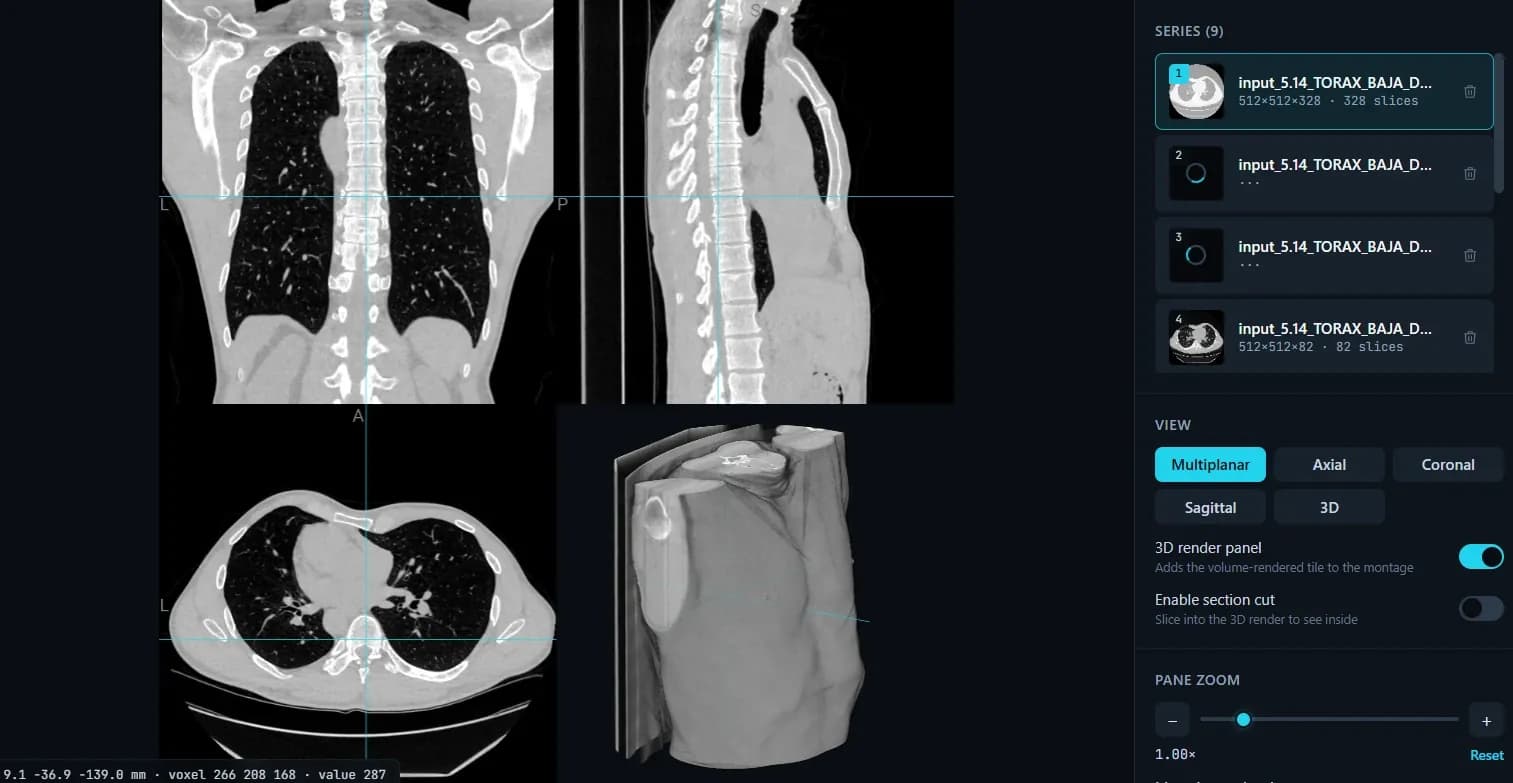
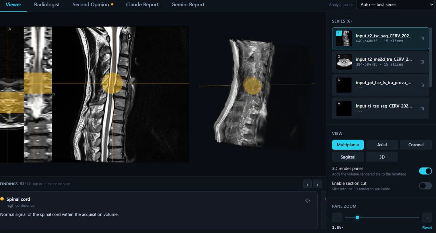
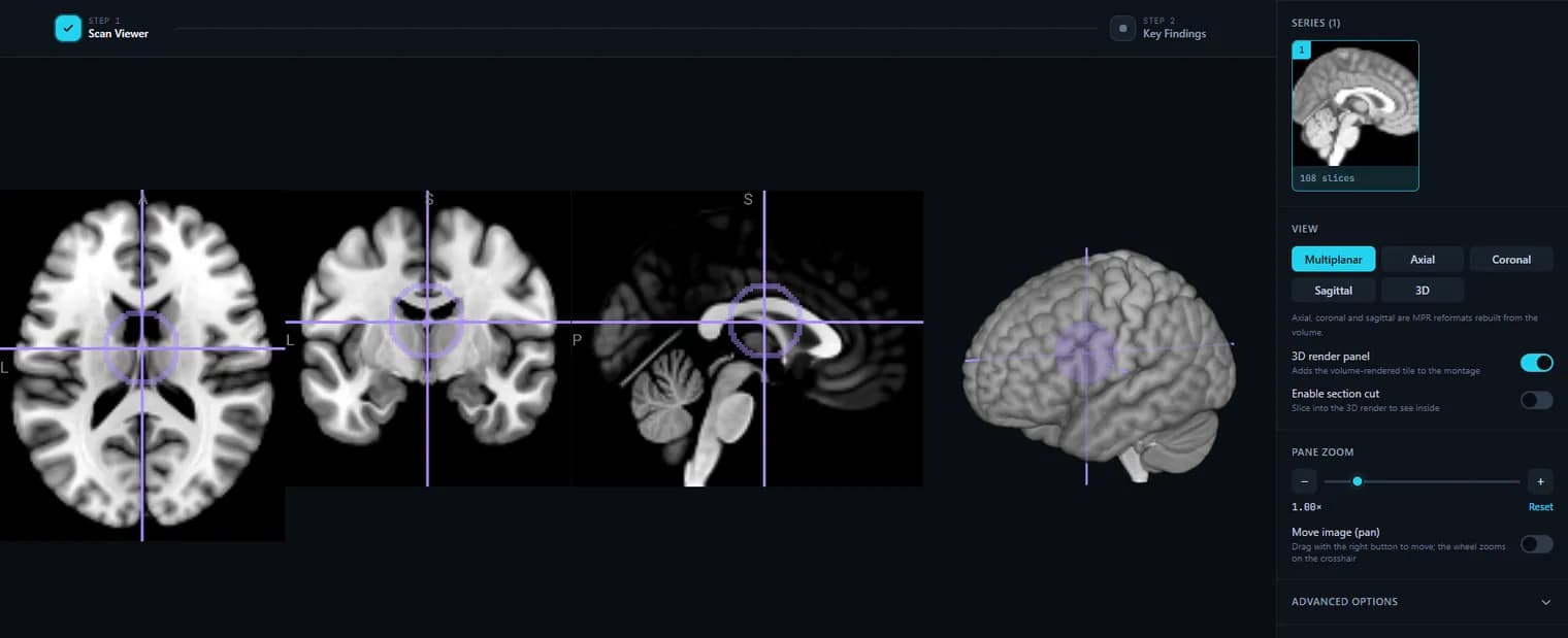
Opens: MRI as a DICOM folder (.dcm) or a NIfTI file (.nii / .nii.gz). · Files are processed in your browser — nothing is uploaded.
What this tool does
Opens the CD, DVD or USB stick the hospital gave you
Insert the disc, choose the drive (or the DICOM folder on the stick), and the study opens. It works the same on Windows and on a Mac — the usual sticking point, because the viewer software burned onto these discs is almost always a Windows .exe that a Mac cannot run at all.
Nothing to install and no account
This is a web page, not a download. There is no installer to talk your IT department into, no licence, and no sign-up before you see your images — which also means nothing lands on a work laptop you are not allowed to install software on.
Every sequence in the study, side by side
An MRI is not one image set: T1, T2, FLAIR, STIR, DWI and post-contrast series each arrive separately. All of them appear in the series list with descriptions and slice counts, so you can move between the sequences your report refers to instead of guessing which one you are looking at.
Scroll the stack the way the radiologist does
Mouse wheel through the slices, arrow keys for one at a time (Shift for ten), the scrubber bar to jump, or a finger swipe on a tablet. The slice number and the total are always on screen, so a report that says "series 4, image 17" becomes something you can actually find.
Axial, coronal and sagittal from one acquisition
The viewer rebuilds the volume, so it can reformat planes the scan was not acquired in, with a shared crosshair linking them. One honest limit: reformatting a thick-slice 2D sequence gives a coarse, stair-stepped second plane — that is the data, not the viewer, and the smoothing toggle lets you see it plainly.
3D rendering with a section cut
Add the 3D tile, rotate it, and switch on the section cut to slice the render open and look inside. It works best on 3D isotropic sequences (MPRAGE, SPACE, CUBE); a five-millimetre 2D stack has too few slices to render convincingly.
Contrast you control
Drag on the image to window it live, or set an exact min/max intensity range, and choose from six colormaps. MRI intensities are not calibrated units the way CT Hounsfield numbers are, so windowing is how you make a subtle signal change visible.
Send one slice, or take the whole volume with you
Save the current view as a PNG for an email or a message, or export the volume as a de-identified NIfTI (.nii.gz) that any research or open-source tool will open.
Your scan stays on your device
The DICOM files are decoded in your browser by dcm2niix compiled to WebAssembly. Using the free viewer uploads nothing — there is no copy of your MRI on a server for anyone to have to trust.
What it does not do
It shows the images; it does not tell you whether they are normal, and it has no measurement, ROI or annotation tools. It is not a diagnostic-grade display and does not replace your radiologist. A disc stored in a compressed transfer syntax we cannot decode in a browser (JPEG 2000 is the usual one) gives you a clear message and the next step, which is desktop dcm2niix.
How to use it
- 1.Upload your MRI DICOM folder or a NIfTI file.
- 2.Scroll through slices and switch between axial, coronal and sagittal planes.
- 3.Use the 3D view and intensity window to inspect the anatomy.
- 4.Want it explained? Create an account for a plain-language AI report.
FAQ
- Can I view my MRI from a hospital CD?
- Yes. Point the “Upload DICOM folder” picker at the DICOM folder on the CD.
- Will this tell me if my MRI is normal?
- The free viewer shows the images only. An optional AI report explains findings in plain language, but it is informational and not a diagnosis — always discuss results with your doctor.
- Is it private?
- Yes — MRI images are processed in your browser and are not uploaded when you use the free viewer.
- Can I open my MRI scan CD or DVD?
- Yes. Insert the disc, click “DICOM folder / CD” and choose the CD/DVD drive — your MRI opens right here in your browser, on Windows or Mac, with nothing to install. Your files are never uploaded. If the disc stores its images in a compressed format we can’t read yet (for example JPEG 2000), you’ll get a clear message telling you the next step.
- Does it support MPR and MIP?
- Yes. The viewer reads the whole volume rather than a stack of pictures, so the axial, coronal and sagittal views are genuine MPR reformats. Under Advanced options you can switch on a slab projection and set its thickness in millimetres: MIP keeps the brightest voxel through the slab (vessels, bone), MinIP the darkest (air, airways). Projections work on one plane at a time; oblique and curved (CPR) reformats are not supported yet.
Want your scan explained in plain language?
The viewer is free — and so is understanding your radiologist’s report. Create an account, upload your scan (de-identified in your browser) and your report, and every finding is mapped onto the images, explained in plain language and scored — no card needed. An independent AI reading of the images is a paid extra. Informational only — not a medical diagnosis.