CT · Chest
What a chest CT shows, and what the report means
Read Your Scan Editorial Team·Last updated
Written from published radiology references and reviewed for plain language. Not written or signed by a physician, and not a diagnosis. How we write these pages →
The short answer
A chest CT shows the lungs and airways, the mediastinum between them, lymph nodes, blood vessels, the heart’s outline and the chest wall, in thin cross-sectional slices. It is ordered to look more closely at a cough, breathlessness, an abnormal X-ray, a known nodule, or an infection.
A chest CT (computed tomography) uses X-rays to build detailed cross-sectional images of your lungs, airways, and the structures around them. This page explains, in plain words, what the scan looks at and what terms on the report can mean.
What this scan shows
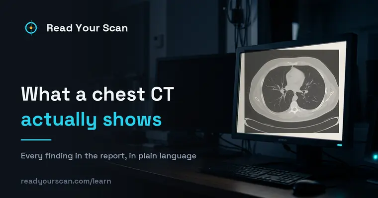
A chest CT gives a much more detailed view than a regular chest X-ray. It shows the lungs and airways, the space between the lungs (the mediastinum), lymph nodes, blood vessels, the heart’s outline, and the chest wall. Doctors order it to look more closely at a cough, shortness of breath, an abnormal X-ray, a known nodule, an infection, or to check blood vessels. Seeing something on the images does not by itself mean something is wrong — your doctor reads the pictures together with your history and other tests.

Common findings, in plain words
Pulmonary nodule
A small round spot in the lung. Nodules are very common and most are harmless — scar tissue or a past infection. Reports often note the size; small ones are frequently just watched over time. Your doctor will decide whether it needs any follow-up.
Ground-glass opacity
A hazy area where the lung looks slightly cloudy rather than solid. It can appear with infections, inflammation, or fluid, and often clears up. It is a description of how the tissue looks, not a diagnosis on its own.
Pleural effusion
A collection of fluid in the space around the lung. It can happen for many reasons, from infection to heart or other conditions. Your doctor will consider why the fluid is there.
Emphysema
Areas where the tiny air sacs of the lung are enlarged or damaged, often linked to smoking. Reports may describe it as mild, moderate, or severe. It describes lung structure and is interpreted alongside your breathing and history.
Enlarged lymph node
A lymph node that looks bigger than usual. Nodes commonly enlarge in response to infection or inflammation and often return to normal. Size is one clue your doctor weighs among many.
Coronary artery calcification
Calcium seen in the walls of the heart’s arteries, sometimes noted incidentally. It is a marker your doctor may discuss with you in the context of heart-health risk.
Pleural parenchymal scarring
Fibrous tissue where the lung meets its lining, left behind by an old infection or inflammation that has healed. It is usually stable and often noted only for comparison with future scans.
Terms you might see
- Contrast
- A dye given through a vein that makes blood vessels and some tissues show up more clearly on the images. Not every chest CT uses it.
- Mediastinum
- The central area of the chest between the two lungs, containing the heart, major blood vessels, windpipe, and lymph nodes.
- Opacity
- A general word for any area that appears denser or cloudier than normal lung on the scan. It describes appearance, not cause.
- Pleura
- The thin lining around the lungs and inside the chest wall. Fluid or thickening here is described as pleural.
- Incidental finding
- Something noticed by chance that was not the reason for the scan. Many are harmless, and your doctor decides if any need attention.
- Air bronchogram
- Air-filled airways showing up as dark branches against lung that has become denser around them. It is a sign that the air sacs, not the airways, are filled with something.
Questions to ask your doctor
- 1.What was the main reason this chest CT was ordered, and did it answer that question?
- 2.Were any findings unexpected, and do any of them need follow-up?
- 3.If a nodule was seen, does it need another scan later, and when?
- 4.Do the results change anything about my current treatment or medications?
- 5.Is there anything in my history that helps explain what the report describes?
See these findings on your own scan
Upload your scan (de-identified in your browser) and your radiologist’s report, and every finding is mapped onto the images, explained in plain language and scored — free, no card needed. Informational only — not a diagnosis.
FAQ
- Does a spot or nodule on my chest CT mean cancer?
- No. Most lung nodules are harmless, such as old scars or the remains of a past infection. Reports simply note them so your doctor can decide whether any watching or follow-up makes sense for you.
- Is a chest CT better than a chest X-ray?
- They serve different purposes. A CT shows much finer detail and cross-sectional slices, while an X-ray is quicker and uses less radiation. Your doctor chooses based on what they need to see.
- What does contrast do, and did I need it?
- Contrast is a dye that highlights blood vessels and certain tissues. Whether it is used depends on the clinical question. Your report or care team can tell you if contrast was part of your scan.
Related
Go deeper
Sources
- Patient-facing procedure descriptions (MRI, CT, X-ray, ultrasound)RadiologyInfo.org — Radiological Society of North America & American College of Radiology
- Guidelines for Management of Incidental Pulmonary Nodules Detected on CT ImagesFleischner Society, via Radiology (RSNA)
- CT scan — overview, how it is performed and resultsNHS (United Kingdom)
Read Your Scan is informational only — not a medical diagnosis, and not a substitute for a licensed radiologist or your doctor. If you have urgent symptoms, seek care.