Understand your scan
What each scan shows, how a radiologist reads it, and what every word on the report means — in plain language, from published references, with the limits stated. Informational only, never a diagnosis.
Written from published radiology references and reviewed for plain language. Not written or signed by a physician, and not a diagnosis. How we write these pages →
Report glossary, A–ZEvery word that shows up on an MRI, CT, X-ray or ultrasound report, explained in plain language.In depth
Longer reads: how a radiologist actually works through a scan, what normal looks like, and the questions the modality guides below deliberately leave to a separate page.
- How to read a radiology reportA radiology report has five parts and only one is the answer. What each section means, which sentence to read first, and what the hedging words are doing.
- MRI vs CT scan: what each one showsMRI and CT answer different questions. What each physically detects, which tissues it sees well, how long it takes, and why your doctor chose one.
- How to read a brain CT scanWhat a radiologist checks on a head CT and in what order: windows, symmetry, grey-white contrast, ventricles, cisterns — and what an early scan misses.
- How to read a chest CT scanLung window, mediastinal window, and the order a radiologist searches a chest CT — what normal lung looks like, and what a nodule or effusion changes.
- How to read a brain MRIA brain MRI is several scans of the same head. What T1, T2, FLAIR, DWI and post-contrast each reveal, and how a radiologist compares them.
- How to read a knee MRIThe three planes, the fluid-sensitive sequences, and the checklist a radiologist works through: meniscus, ligaments, cartilage, bone marrow.
- How to read a lumbar spine MRILevels, discs and nerve roots: what bulge, protrusion, extrusion, stenosis and foraminal narrowing actually describe — and what a lumbar MRI does not cover.
- MRI sequences explainedWhy one MRI produces many different image sets, what each sequence is sensitive to, and how to tell fat from fluid from blood by comparing them.
- Contrast on a CT or MRI, explainedWhat contrast dye does, why iodine is used for CT and gadolinium for MRI, why some scans are done both with and without, and what the safety checks are for.
- How long do scan results take?The images are ready in minutes; the report is not. Why routine scans wait, what urgent changes, and why the portal shows it before your doctor calls.
MRI
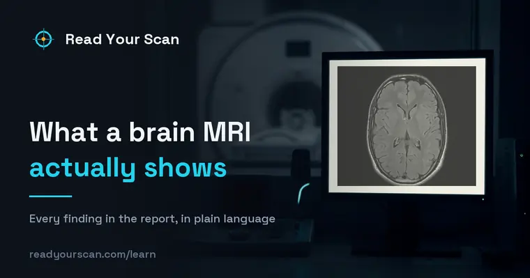 What a brain MRI shows, and what the report meansA brain MRI shows brain tissue, fluid spaces and vessels in fine detail, without radiation. What the common findings and terms on the report mean.
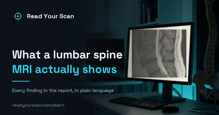 What a lumbar spine MRI shows, and what the report meansA lumbar MRI shows the discs, spinal canal, nerve roots and facet joints, level by level. What bulge, stenosis and degenerative change mean.
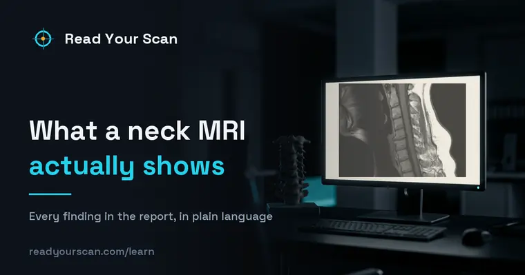 What a cervical spine MRI shows, and what the report meansA cervical MRI shows the neck vertebrae, the discs, the spinal cord and the nerve roots. What the common findings on a neck MRI report mean.
 What a knee MRI shows, and what the report meansA knee MRI shows the menisci, ligaments, cartilage, bone marrow and joint fluid. What a tear, an effusion and bone oedema mean on the report.
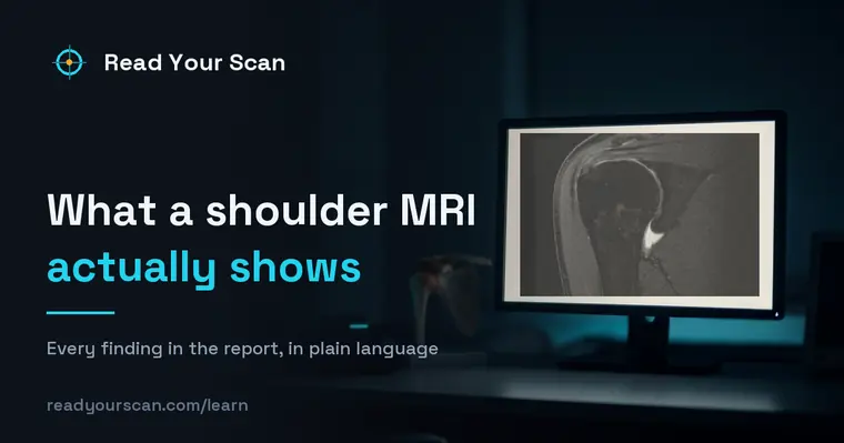 What a shoulder MRI shows, and what the report meansA shoulder MRI shows the rotator cuff, labrum, capsule, cartilage and bone marrow. What a tear, tendinopathy and impingement mean on the report.
CT
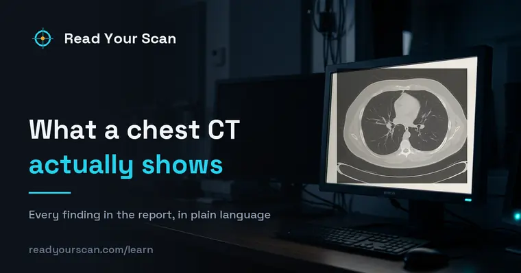 What a chest CT shows, and what the report meansA chest CT shows the lungs, airways, lymph nodes and blood vessels in cross-section. What each common finding on the report means, in plain words.
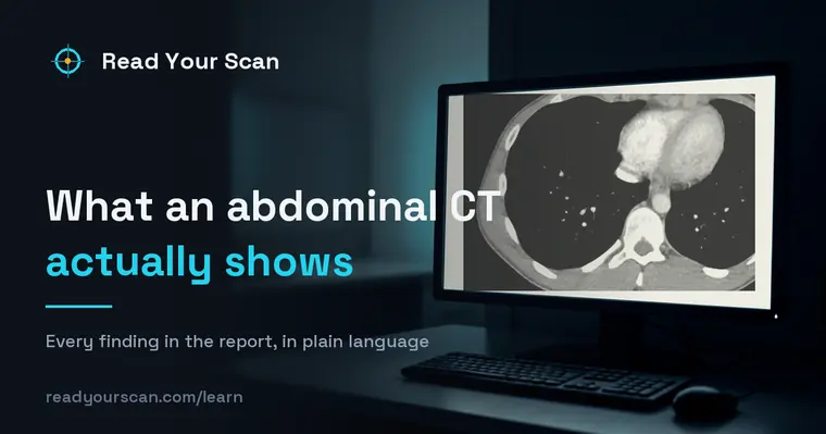 What an abdominal CT shows, and what the report meansAn abdominal CT shows the liver, kidneys, pancreas, spleen, bowel and vessels in cross-section. What the common findings on the report mean.
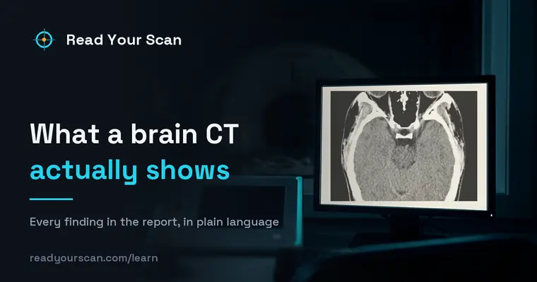 What a brain CT shows, and what the report meansA head CT shows bleeding, the fluid spaces, the skull and gross brain structure in a minute. What it detects, what it misses, and what it means.
X-ray
Ultrasound
From the blog
Blog home →One article per report phrase or per finding — what it means, how common it is, and what usually happens next. Every image is a real, openly licensed MRI, CT or X-ray.
Report language
- What “clinical correlation is recommended” means on your reportThe sentence is addressed to your doctor, not to you. What a radiologist means by it, the three different jobs it does, and when it is worth asking about.
- What “lucency” means on an X-ray or CT reportLucency means an area let more X-rays through — it looks darker on the film. What that describes, why it is a shape and not a diagnosis, and what changes its meaning.
- What “unremarkable” means on a medical reportUnremarkable means normal — nothing worth remarking on. Why the best news in a radiology report is written in its flattest word, and what "grossly" adds.
- What “effacement of the thecal sac” means on a spine MRIThe thecal sac is the fluid-filled sleeve around the spinal nerves. Effacement means something is pressing on its edge — what that does and does not imply.
- What “the cardiomediastinal silhouette is unremarkable” meansThe longest word on a normal chest X-ray report, and one of the best. What the cardiomediastinal silhouette is and why its outline is what gets described.
- What “no acute osseous abnormality” meansThe standard sentence for "no broken bone" — and what the word "acute" is quietly leaving room for. What the phrase covers and what it does not.
- What “no acute intracranial abnormality” means on a head CTThe standard conclusion of a normal head CT. What the scan was actually looking for, why "acute" is the key word, and what a normal CT does not rule out.
Findings explained
- What “small vessel ischemic changes” means on a brain MRIWhite spots in the brain’s white matter, reported as small vessel disease or chronic microvascular change. Why they are so common after 50 and what the wording is actually claiming.
- What “straightening of the cervical lordosis” meansYour neck X-ray or MRI says the normal curve is straightened or reversed. What that describes, why muscle spasm and the scan position cause it, and why the finding alone rarely explains pain.
- What an “adrenal incidentaloma” is on a CT reportA lump on an adrenal gland, found on a scan done for something else. Why most are harmless, what Hounsfield units have to do with it, and the two questions that decide what happens next.
- What “retrolisthesis” means on a spine reportOne vertebra sitting slightly backwards on the one below. What the grades mean, why it usually comes from disc wear rather than injury, and when it matters.
- What a “Tarlov cyst” is on a spine MRIA fluid-filled pouch on a nerve root, almost always at the base of the spine. Why most are found by accident and cause nothing, and why the symptomatic minority is genuinely contested.
- What “bone marrow edema” means on an MRIFluid inside a bone, seen only on MRI. Why it is a signal rather than a diagnosis, what a bone bruise really is, and why the same words cover an injury and arthritis.
- What “AC joint osteoarthritis” means on a shoulder reportWear at the small joint where the collarbone meets the shoulder blade. Why almost every adult shoulder scan mentions it, and how to tell whether it is the thing causing your pain.
- What “patellar tendinosis” means on a knee MRIWhy the report says tendinosis and not tendinitis, what that changes about treatment, and why the tendon looks abnormal in people with no pain.
- What “subacromial-subdeltoid bursitis” meansFluid in the cushion above the rotator cuff. What the report is measuring, why the bursa is almost never inflamed on its own, and what usually sits underneath it.
- What “subchondral sclerosis” means on a reportBone under the cartilage looking whiter and denser. Why it is the bone responding to load rather than a disease of its own, and what it sits alongside.
- What “concha bullosa” means on a sinus CTAn air pocket inside one of the structures in your nose. Why it is classed as a normal variant found in a third of people, and when it is worth doing anything about.
- T2/FLAIR hyperintensities: the bright spots on a brain MRIThe bright spots on your brain MRI, what the report calls them, why "small vessel ischemic disease" sounds worse than it usually is, and how they are graded.
- Modic changes type 1, 2 and 3 on a spine MRIWhat Modic changes are, what the three types actually describe, why type 1 is the one associated with pain, and what the grading does and does not predict.
- What “mucosal thickening” means on a sinus CT or MRIWhy "mucosal thickening" turns up on scans done for something else entirely, what mild versus severe means, and when a sinus finding is worth acting on.
- Schmorl’s nodes: what they are on a spine reportA Schmorl node is disc material pressed into the bone of a vertebra. Why they are usually old, usually painless, and why so many reports mention them.
- What “joint effusion” means on an MRI reportExtra fluid in a joint. Why a small effusion is normal, what "trace" and "moderate" are measuring, and why the effusion is a clue rather than the problem.
- What “ground-glass opacity” means on a lung CTA hazy area on a lung CT that does not hide the structures underneath. What causes it, why "ground glass" is a description and not a diagnosis, and what follows.
- What “facet arthropathy” and “facet hypertrophy” meanArthritis of the small joints at the back of the spine. What hypertrophy is describing, why it is nearly universal after middle age, and when it explains pain.
- What “encephalomalacia” means on a brain scanAn area where brain tissue was lost and never came back. Why it is almost always old, what gliosis beside it means, and why it is not a progressive disease.
- What phleboliths are on a CT or X-ray reportSmall calcium stones inside pelvic veins. Why nearly half of adults have them, why they are harmless, and why they matter when someone is looking for a kidney stone.
- What a pineal cyst means on a brain MRIA fluid-filled sac in a gland at the centre of the brain. Why most are found by accident, why size alone is not the issue, and when one is actually followed up.
- What “cavum septum pellucidum” means on a brain scanA small fluid space between two membranes at the centre of the brain. Why it is a normal variant in adults, and why it is looked for on a fetal scan.
Ready to understand your own images? Get your report explained free or open the free viewer.