Your radiologist read it once.
Your medical scan.
Analyzed by AI.
Upload your MRI, CT, X-ray or ultrasound. The AI reads every image without seeing your report: an AI-assisted Second Opinion in plain words, mapped on your images. Add it to see where the two agree.
Drop your scan or report here
DICOM, images (JPG/PNG) or a report PDF — add it all together
- Encrypted in transit & at rest
- You control deletion
- We never sell your data
- Results in minutes, not days
- Report explained free — no card
- 3D viewer free, no account
- Scored normal → significant
- Not a diagnosis
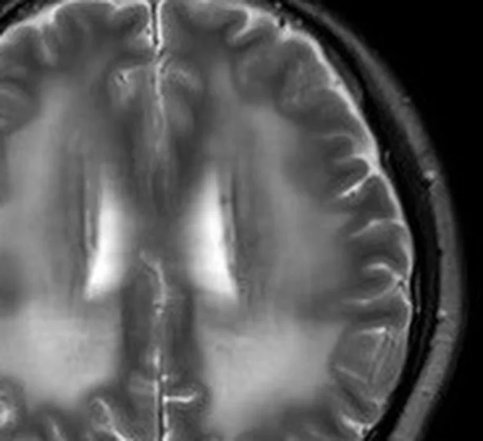
MRI · brainNormal
“Multiple T2-hyperintense foci in the periventricular white matter, of probable microvascular origin.”
→ A few small bright spots in the brain’s wiring, next to the fluid spaces. Very common with age — on their own, not a disease.
real, openly-licensed MRI and CT exams — example findings, not a diagnosis · credits
How it works
One scan, three steps.
The same exam all the way through: it goes in, your radiologist’s report gets pinned onto it, and it comes back with a second reading.
- DICOM folder / CDNIfTIX-ray photo
Series
 1
1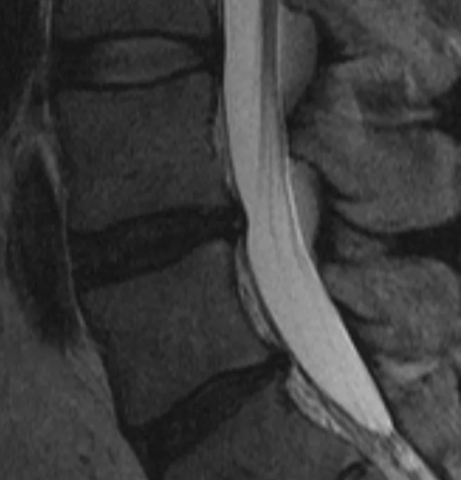 2
2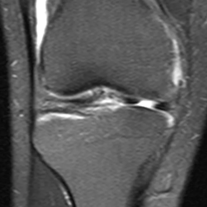 3
3De-identified in your browser
PatientNamePatientIDBirthDate→ removed
01Upload your scan
A DICOM folder or the hospital CD, a NIfTI file, or a photo of an X-ray. Name, ID and dates are stripped in your browser before anything is uploaded.
Free · the viewer needs no account
Report · PDF or pasted text
Conus medullaris terminates at L1. Vertebral body heights are preserved.
Reduced height and T2 signal of the L5–S1 disc, with a broad posterior bulge indenting the thecal sac.
No spondylolisthesis. Paraspinal soft tissues unremarkable.
L5–S1Significant
→ The lowest disc in the back has dried out and lost height, and now pushes back onto the sac of fluid around the nerves.
02Add the radiologist’s report
Paste the text or upload the PDF. Every sentence becomes a finding: rewritten in plain words, scored, and pinned to the exact spot on your images.
Free · no card
- MRI · lumbar spineYour radiologistAI second read
- L5–S1 disc bulge onto the thecal sac✓✓both
- Reduced height of the L5–S1 disc✓✓both
- Early drying of the L4–L5 disc—✓AI only
Raised by one reader only — bring it to your appointment.
03Get an AI Second Opinion
Deep Analysis reads every series from scratch, without seeing what your radiologist wrote. Where the two agree, read with more confidence; what only one raises is what to bring to your appointment.
$9 once · never expires · not a diagnosis
Real, openly-licensed lumbar MRI. The report sentence and the comparison are representative examples, not the source exam’s own report — and not a diagnosis.
Want to see a whole reading first? Open the example — no upload needed.
See a sample AI reading →Features
See it. Understand it. Share it.
Everything happens on your own images — never on a summary of them.
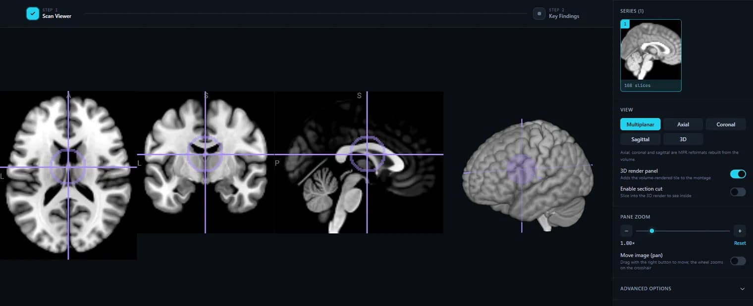
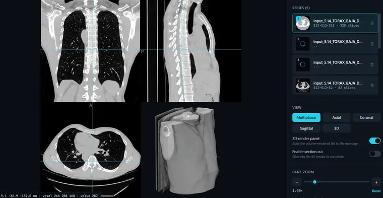 Screenshot · the actual viewer
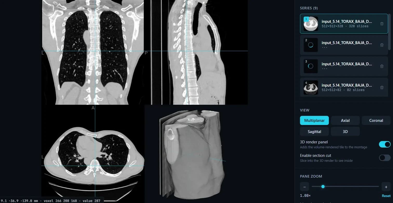
A real 3D viewer, in the browser
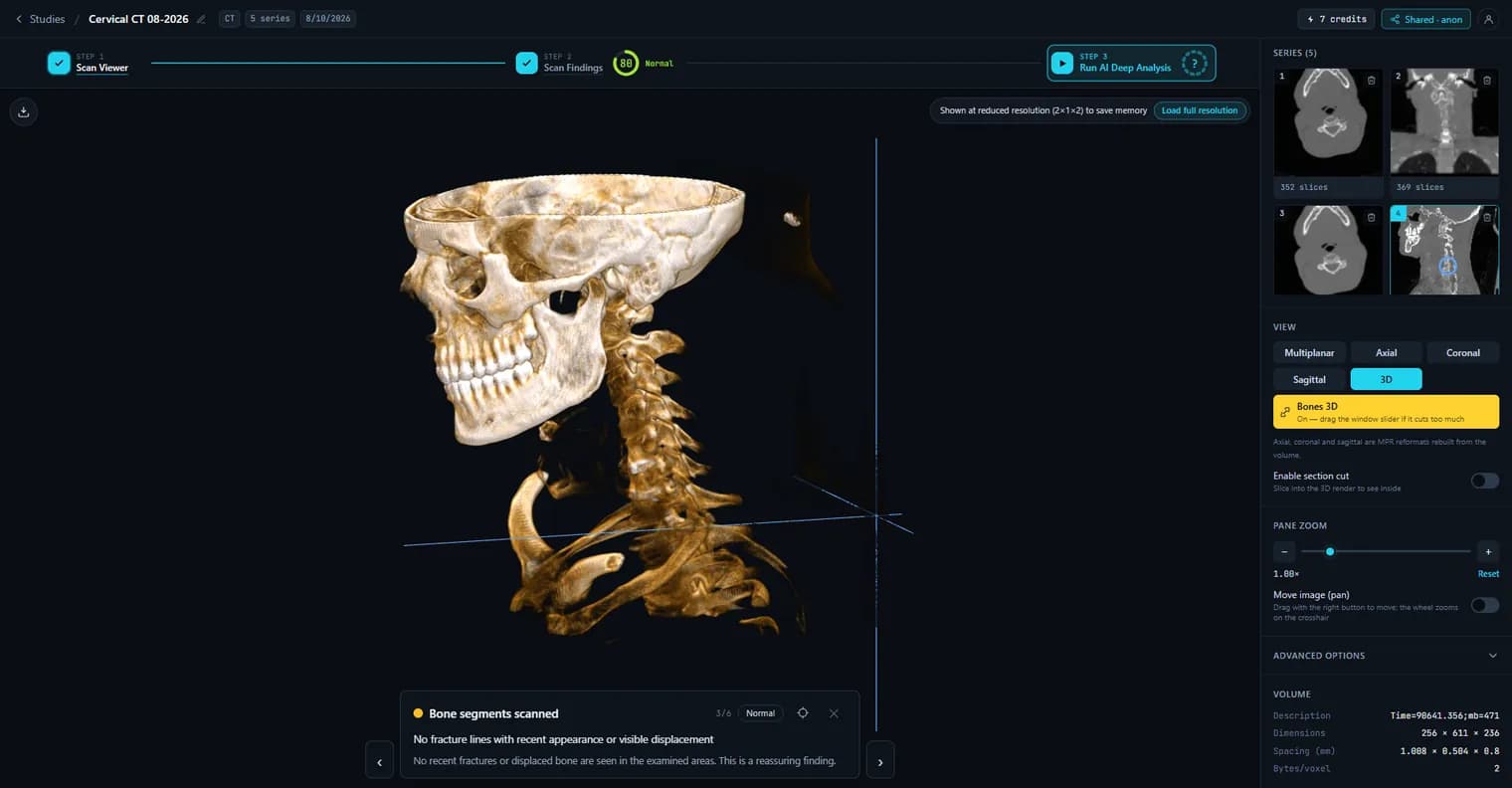
Axial, coronal, sagittal and a volume render of MRI, CT and X-ray — nothing to install, no account for the viewer.
Open the free viewer →Every finding, pinned on the image
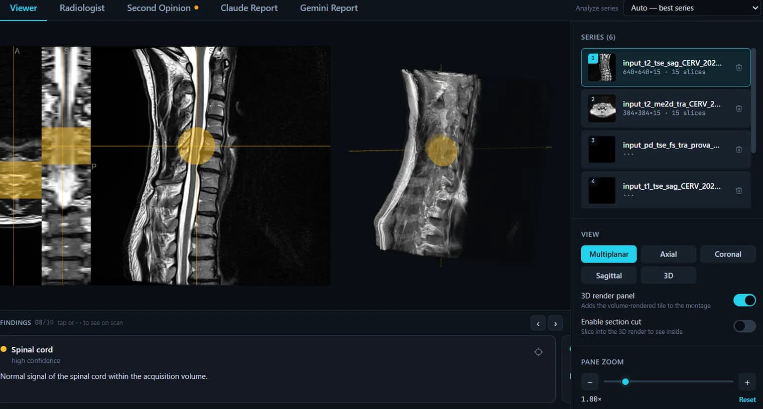
Click a finding and the viewer jumps to the exact spot it describes — the words on the page become regions you can see.
Report explained →- 3 normal
- 2 needs attention
- 1 significant
Scored, normal → significant
Each finding gets a severity that echoes the report’s own wording, and the whole exam rolls up into one score. Never a new judgment.
How the score works →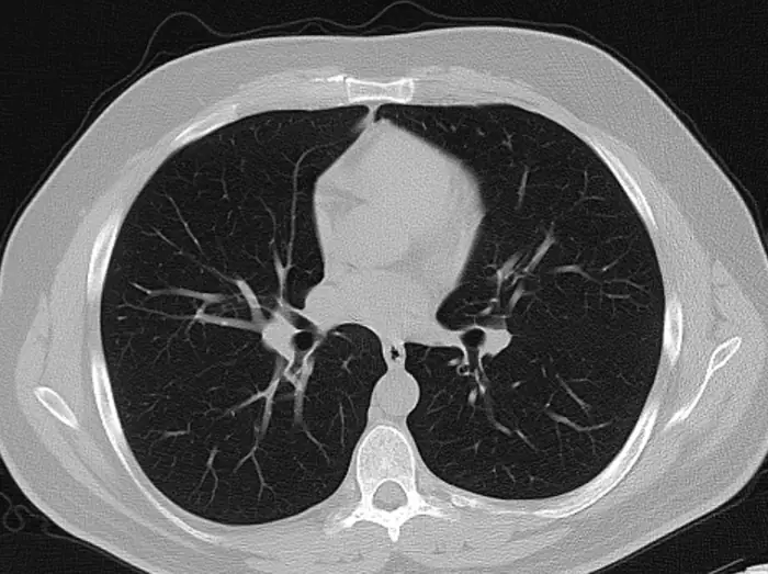
An AI reading that never saw the report
Deep Analysis reads every series of the exam from scratch. Put it beside your radiologist’s: agreement reassures, a difference is a question worth asking.
AI second opinion →Is the L5–S1 bulge pressing on a nerve?
Your report says it indents the thecal sac and does not mention a nerve root. Whether it explains your leg symptoms is a question for your doctor.
Ask follow-up questions
Answered from your own report and images, not from the internet. Informational — the answer always points you back to your doctor.
AI radiology report →- no account for your doctor
- read-only
- revoke any time
Send it to your doctor in one link
Images plus the mapped findings, on a private link they open without an account — or a read-only public one. Revoke any time.
Share by link →Severity at a glance
“How bad is it?” — answered first.
Every AI reading opens with a severity summary: a gauge that rolls up the whole study, a health score — the same 0–100% you’ll see on your dashboard — and counts per bucket: normal, needs attention, significant. When the score drops into the amber or red band, that’s a clear signal to book a follow-up with your doctor — and these findings are exactly what to bring along. The score echoes the report’s own wording; it never invents a new clinical judgment.
Key findings · MRI cervical spine · 5 findings
A score in this band is a clear indication to follow up with your doctor. Severity reflects the wording of the report itself. Informational — not a diagnosis. If you feel unwell, contact your doctor or urgent care.
Deep Analysis
Your whole exam, read series by series.
Not a chatbot glancing at a screenshot. A general chatbot like ChatGPT, Grok, Claude or Gemini comments on a flat photo — it can’t open your real DICOM volume or scroll the slices. Deep Analysis reads your actual study series by series, working from the scan’s own geometry, then turns what it saw into a plain-language report.
Reads the real thing
Your real volume, not a photo
A chatbot sees one compressed screenshot. Deep Analysis works from your actual study — slices taken across every diagnostic series of the exam.
Reads it in context
It knows what it is looking at
Which plane each slice is, and which side is your right, are read from the scan’s own geometry rather than guessed from the picture — and the reading knows your age, sex and the symptoms you described.
Answers you can use
Mapped, scored, ready for your doctor
Every finding lands on your images with a severity score — plus follow-up questions grounded in your study.
The readers
Frontier models, picked by measurement
A chatbot hands you whatever model it happens to run. We keep several and let measurement decide: readers are compared head-to-head on real studies with a known diagnosis, and the one that wins reads yours — which is why a flat X-ray and a volumetric MRI don’t go to the same model. The roster includes models built for medical imaging that no chatbot can reach, such as Google’s MedGemma.
- Claude Opus
- Gemini
- MedGemma 1.5
How it compares
ChatGPT is a generalist. Read Your Scan is built for medical imaging.
Both explain a report or comment on a photo. Only one opens your real exam and reads every slice of it.
| ChatGPT | Read Your Scan | |
|---|---|---|
| Explains the report in plain words | ||
| Comments on a single uploaded image | ||
| Reads the scan’s own geometry — the plane, and which side is your right (Deep Analysis) * | ||
| Opens the real 3D volume from DICOM / NIfTI, not a screenshot | ||
| Analyzes the whole exam — every series and slice | ||
| Findings anchored to anatomy, clickable on the images | ||
| De-identified in your browser before anything is uploaded (GDPR) | ||
| Stores and organizes your exams over time | ||
| Share a 3D study with a doctor |
* Deep Analysis is a paid feature.
“ChatGPT” stands in for general chatbots (also Claude, Gemini, Grok). Every tool here is informational, not a medical diagnosis.
The real viewer
This is what your scan looks like here
CD or DVD?
Got your scan on a hospital CD?
Insert the disc and open your MRI, CT, X-ray or ultrasound right in your browser — nothing to install, works on Mac and Windows, and your files are never uploaded.
Your scan archive
All your scans, in one place online.
Got a drawer full of hospital CDs and DVDs? Copy each exam into your account and keep your whole imaging history in one secure place — instead of discs that scratch, get lost, or won’t open on a modern computer.
- Import from any scan CD or DVD
- Open any exam in 3D, anytime
- Share any one with a doctor by link
Your exams stay for as long as you use your account. If an account goes unused for six months, an exam that never had a reading may be deleted — and we email you before that happens. Data retention
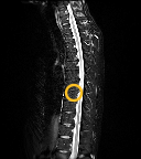
Understand your scan
Plain-language guides to what each scan shows
Not sure what a finding on your report means? These free guides explain MRI, CT, X-ray and ultrasound in plain words — the common findings and the jargon — so you can read your own imaging. Browse all guides →
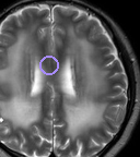
MRI · Brain
What a brain MRI shows, and what the report means
A brain MRI shows brain tissue, fluid spaces and vessels in fine detail, without radiation. What the common findings and terms on the report mean.
CT · Chest
What a chest CT shows, and what the report means
A chest CT shows the lungs, airways, lymph nodes and blood vessels in cross-section. What each common finding on the report means, in plain words.
CT · Brain
What a brain CT shows, and what the report means
A head CT shows bleeding, the fluid spaces, the skull and gross brain structure in a minute. What it detects, what it misses, and what it means.
X-ray · Chest
What a chest X-ray shows, and what the report means
A chest X-ray shows the lungs, heart outline, ribs and diaphragm in one projection. What consolidation, effusion and cardiomegaly describe.
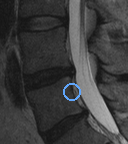
MRI · Spine
What a lumbar spine MRI shows, and what the report means
A lumbar MRI shows the discs, spinal canal, nerve roots and facet joints, level by level. What bulge, stenosis and degenerative change mean.
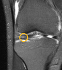
MRI · Knee
What a knee MRI shows, and what the report means
A knee MRI shows the menisci, ligaments, cartilage, bone marrow and joint fluid. What a tear, an effusion and bone oedema mean on the report.
A–Z
Report glossary, A–Z
Every word that shows up on an MRI, CT, X-ray or ultrasound report, explained in plain language.
From the blog
One report phrase, one finding, one article Read the blog →
What “small vessel ischemic changes” means on a brain MRI
White spots in the brain’s white matter, reported as small vessel disease or chronic microvascular change. Why they are so common after 50 and what the wording is actually claiming.
What “straightening of the cervical lordosis” means
Your neck X-ray or MRI says the normal curve is straightened or reversed. What that describes, why muscle spasm and the scan position cause it, and why the finding alone rarely explains pain.
What an “adrenal incidentaloma” is on a CT report
A lump on an adrenal gland, found on a scan done for something else. Why most are harmless, what Hounsfield units have to do with it, and the two questions that decide what happens next.
What a BI-RADS 3 result means
Probably benign, with a short-interval follow-up in six months. What the category actually claims, the number behind "probably", and why the waiting is the hard part.
What “retrolisthesis” means on a spine report
One vertebra sitting slightly backwards on the one below. What the grades mean, why it usually comes from disc wear rather than injury, and when it matters.
What a “Tarlov cyst” is on a spine MRI
A fluid-filled pouch on a nerve root, almost always at the base of the spine. Why most are found by accident and cause nothing, and why the symptomatic minority is genuinely contested.
Sharing
Send it to your doctor in one link
Share medical images by link — privately with your doctor, or read-only in public.
Private link
Share a study — images plus the mapped findings — with your doctor via a private link only they can open. No account needed on their side.
Public link
Or publish a read-only public link when you want to share more widely. You stay in control — revoke access at any time.
De-identified by design
Identifying DICOM tags — name, IDs, dates — are stripped in your browser before anything is uploaded, so a shared link shows the images and findings, never your personal details.
Any language
Read your report in your language.
Translate the plain-language report and findings into the major world languages with AI — the medical terms stay accurate, the explanation stays clear.
Privacy
Your data stays yours.
This is medical data, and we treat it that way. The riskiest thing is your identity — so we strip it before your scan ever leaves your device.
- Imaging is de-identified in your browser before anything is uploaded.
- Encrypted in transit and at rest. We never sell or share your medical data.
- Your studies stay private to your account — share, export or delete anytime.
- An EU company — your scans handled under GDPR health-data rules.
Pricing
The viewer is free. You pay per exam, once.
Viewing scans, and having your radiologist’s report mapped, explained and scored, are always free. You pay only for an independent AI reading of the images — once per exam, no subscription, nothing expires.
3D viewer and sharing, plus your radiologist’s report mapped onto your scan, explained in plain language and scored — all free when you sign up, no card needed.
- Read in full for free: severity score and finding count shown
- Unlock the written report — the AI reading of your pictures
- For flat pictures, not a 3D MRI/CT volume
- Every series in the exam, read independently — not a summary of your radiologist’s report
- Findings pinned on your images and scored for severity
- Full plain-language report · PDF
- Read it in 9 languages
- Follow-up questions, answered from your report
For a drawer of old scans, or your family · never expires
Get startedFAQ
Questions, answered
Is this a medical diagnosis?+
No. Read Your Scan gives you an informational, plain-language explanation of your imaging or report, and lets you view it in 3D. It is not a diagnosis and not a substitute for your doctor or a radiologist. Always confirm with your care team.
What makes the viewer different?+
It renders your MRI, CT or X-ray as a real 3D volume in the browser with WebGL — most free online viewers only show flat 2D slices. And every AI or radiologist finding is clickable, jumping the viewer to the exact region.
Can it use my existing radiologist report?+
Yes. Upload the report PDF or paste the text, and each finding is extracted and mapped onto your images, so the written report becomes regions you can see and click.
Can I share a study with my doctor?+
Yes — share a private link only your doctor can open, or a read-only public link when you want to share more widely. You can revoke access at any time.
Can I read it in another language?+
Yes. The report and findings can be translated into the major world languages with AI, keeping the medical terms accurate.
How much does it cost?+
The 3D viewer, sharing, and understanding your radiologist’s report — mapped onto your scan, explained in plain language and scored — are all free, no card needed. A full independent AI reading of the images (Deep Analysis reads the scan itself, beyond your written report, with follow-up questions) is a one-time $9, or $24 for three exams — $8 each — if you have a drawer of old scans or a family to cover. Bought once, never expires, no subscription.
Is my data private?+
Your imaging is de-identified in your browser before upload, encrypted in transit and at rest, and never sold. You can share, export or delete everything at any time.
See your scan the way it should be seen.
Open the free 3D viewer now — or upload a study and let AI read it, in plain language you can click through.
Real, openly-licensed exam slices used on this page — brain: Huang X. et al., Front Neurol 2023, via Wikimedia Commons, CC BY 4.0 · lumbar spine: Stillwaterising, Wikimedia Commons, CC0 1.0 · knee: Ciernik M., Wikimedia Commons, CC BY-SA 4.0 · chest: Ptrump16, Wikimedia Commons, CC BY-SA 4.0. Cropped and marked by us; the knee and chest derivatives are shared under CC BY-SA 4.0.