MRI · Spine
What a lumbar spine MRI shows, and what the report means
Read Your Scan Editorial Team·Last updated
Written from published radiology references and reviewed for plain language. Not written or signed by a physician, and not a diagnosis. How we write these pages →
The short answer
A lumbar spine MRI shows the five lower vertebrae, the discs between them, the spinal canal, the nerve roots leaving at each level, and the facet joints at the back. It does not properly image the hips, the sacroiliac joints or the pelvis, which need their own study.
A lumbar spine MRI makes detailed pictures of your lower back — the bones, discs, and nerves. This page explains, in plain words, what it looks at and what common report terms mean.
What this scan shows
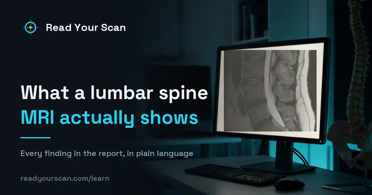
A lumbar (low-back) MRI shows the vertebrae, the cushioning discs between them, the spinal canal, and the nerves that travel to the legs. Doctors order it for low-back pain, sciatica, leg numbness or weakness, or to plan care. Importantly, many of the changes it describes are extremely common and are seen in people with no pain at all — so the report is read together with how you actually feel.

Common findings, in plain words
Disc bulge
A disc that extends a little beyond its normal edge. Bulges are very common and often cause no symptoms. Your doctor considers whether it lines up with what you feel.
Disc herniation
A more focal push of disc material beyond its normal border. Many herniations improve on their own over time. The report notes its location so your doctor can relate it to your symptoms.
Disc desiccation
A disc that has lost some of its water content and appears drier on the images. This is a normal part of aging and is seen in most adults.
Spinal stenosis
A narrowing of the space around the spinal canal or nerves. It can be mild, moderate, or severe. Your doctor interprets the narrowing alongside your symptoms.
Spondylolisthesis
One vertebra sitting slightly forward or backward relative to the one below. It is often mild and stable. The report grades it so your doctor can gauge its significance.
Nerve root compression
A nerve appearing pressed or crowded where it exits the spine. This is described so your doctor can see whether it matches symptoms like leg pain or tingling.
Bone island (enostosis)
A small patch of dense, compact bone sitting inside normal bone, present since childhood in most cases. It is benign, causes nothing, and is reported mainly so it is not mistaken for something else later.
Terms you might see
- Vertebra
- One of the stacked bones that make up the spine. In the low back these are labeled L1 through L5.
- Disc
- The soft cushion between two vertebrae that absorbs shock and allows movement.
- Foramen
- The small opening on each side of the spine where a nerve exits. Narrowing here is called foraminal stenosis.
- Facet joints
- The small joints at the back of the spine that guide movement. Age-related wear here is very common.
- Thecal sac
- The fluid-filled sheath that surrounds the spinal nerves in the canal. Reports often describe whether it is indented.
- Cauda equina
- The bundle of nerve roots that continues down the spinal canal after the spinal cord itself has ended, named for its resemblance to a horse’s tail. It is what lumbar reports are describing when they talk about the nerves.
Questions to ask your doctor
- 1.Do the findings on the MRI match where I actually feel pain or symptoms?
- 2.Are these changes common for my age, or is anything unusual?
- 3.What are my non-surgical options, such as physical therapy?
- 4.Are there any signs that would mean I should seek care sooner?
- 5.Do I need any follow-up imaging, and when?
See these findings on your own scan
Upload your scan (de-identified in your browser) and your radiologist’s report, and every finding is mapped onto the images, explained in plain language and scored — free, no card needed. Informational only — not a diagnosis.
FAQ
- I have a disc bulge — is that serious?
- Disc bulges are extremely common and are found in many people who have no back pain at all. What matters is whether the finding lines up with your symptoms, which is something your doctor interprets for you.
- Does a herniated disc always need surgery?
- No. Many herniated discs improve over weeks to months with non-surgical care. Your doctor considers your symptoms and function, not the images alone, when discussing options.
- Why does my report list so many findings?
- MRI is very detailed and picks up normal age-related changes like disc drying and mild wear. Listing them is thorough reporting, not a list of problems. Your doctor sorts out which, if any, are relevant.
Related
Go deeper
Sources
- Patient-facing procedure descriptions (MRI, CT, X-ray, ultrasound)RadiologyInfo.org — Radiological Society of North America & American College of Radiology
- MRI scan — overview, how it is performed and resultsNHS (United Kingdom)
- ACR Appropriateness Criteria and practice parameters for reportingAmerican College of Radiology
Read Your Scan is informational only — not a medical diagnosis, and not a substitute for a licensed radiologist or your doctor. If you have urgent symptoms, seek care.