CT scan viewer online
Open a CT scan in your browser and inspect it with standard Hounsfield windows — bone, lung, soft tissue and brain. Everything runs locally, so your CT images stay private. Axial, coronal and sagittal views are MPR reformats rebuilt from the volume, and a slab MIP or MinIP is one click away.
Drop your scan or report here
DICOM, images (JPG/PNG) or a report PDF — add it all together
- Encrypted in transit & at rest
- You control deletion
- We never sell your data
- Results in minutes, not days
Opens: CT as a DICOM folder (.dcm) or a NIfTI file (.nii / .nii.gz). · Files are processed in your browser — nothing is uploaded.
What this tool does
One-click Hounsfield window presets
Soft tissue, lung, bone, brain and abdomen presets set the window centre and width the way a CT workstation does. The same structure looks like nothing on the wrong window and obvious on the right one, which is why a single default view of a CT is close to useless.
Or set the window yourself, in HU
Type an exact minimum and maximum, or drag on the image to adjust the contrast live and watch the structure appear. One button resets to the volume’s full range when you have lost your bearings.
The Hounsfield number under the cursor
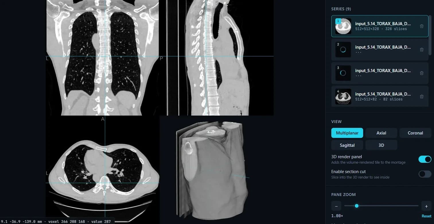
The readout gives world mm, the voxel index and the raw value at that voxel — on a CT, the HU. That number is what separates fat from simple fluid from soft tissue from calcification, and it is the one measurement this viewer can honestly give you.
MIP and MinIP slabs for vessels, bone and airways
Project a slab of a chosen thickness in millimetres rather than one thin slice. Maximum intensity follows a contrast-filled vessel or a bony margin through the slab; minimum intensity brings out air — lung and airway.
Real multiplanar reformats
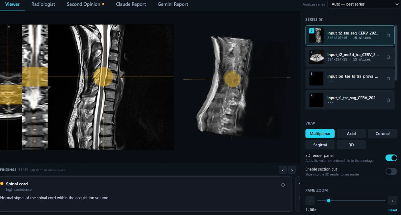
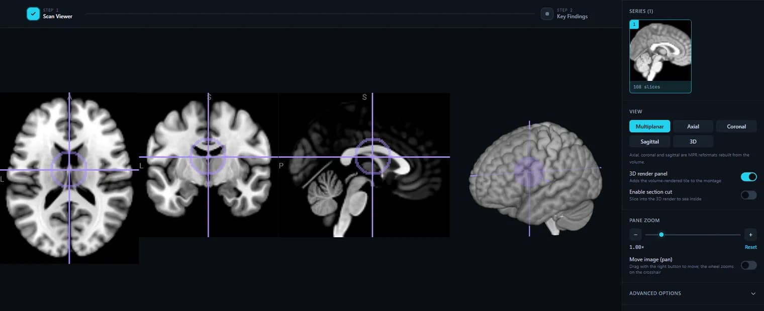
A modern CT is a near-isotropic volume, so coronal and sagittal reformats out of an axial acquisition look genuinely diagnostic rather than smeared. All three planes share one crosshair.
3D volume rendering with a section cut
Rotate the render by azimuth and elevation, set the depth, and clip the volume with the section cut to see inside it. A bone window and a thin-slice CT give the most useful render.
Opens a CT disc on Windows or macOS
Insert the CD or DVD, pick the drive or the DICOM folder, and the study loads in the browser. No installer, no account, and no dependence on the Windows-only viewer burned onto the disc.
Every reconstruction as its own series
A CT study routinely holds several passes — pre- and post-contrast, thin and thick reconstruction, different kernels. They are listed separately with slice counts so you can compare them instead of scrolling through a merged pile.
Large volumes load at reduced resolution first
A whole-body or thin-slice CT can run to hundreds of millions of voxels. Above roughly forty million the volume is box-averaged down so the tab survives, with a labelled button to load it at full resolution when you want the detail.
Nothing is uploaded
DICOM decoding runs in your browser through dcm2niix compiled to WebAssembly. Your CT is not sent anywhere when you use the free viewer.
What it does not do
No distance, angle or ROI measurement; no oblique or curved reformats; no dose or perfusion analysis; no diagnosis. It is not a diagnostic-grade workstation. Discs written in a compressed transfer syntax a browser cannot decode (usually JPEG 2000) are reported clearly and pointed at desktop dcm2niix.
How to use it
- 1.Upload your CT DICOM folder or NIfTI volume.
- 2.Pick a CT window preset (soft tissue, lung, bone, brain) for the right contrast.
- 3.Scroll slices, switch planes and use the 3D render.
FAQ
- Does it have CT windowing (bone/lung/soft tissue)?
- Yes. Load the study as CT and use the one-click Hounsfield window presets in the viewer.
- Are my CT images uploaded?
- No — the free viewer decodes and renders CT locally in your browser.
- Can it explain my CT report?
- You can upload your radiologist report and get a plain-language explanation, and run an AI report on the images — both optional and informational, not a diagnosis.
- Can I open my CT scan CD or DVD?
- Yes. Insert the disc, click “DICOM folder / CD” and choose the CD/DVD drive — your CT scan opens right here in your browser, on Windows or Mac, with nothing to install. Your files are never uploaded. If the disc stores its images in a compressed format we can’t read yet (for example JPEG 2000), you’ll get a clear message telling you the next step.
- Does it support MPR and MIP?
- Yes. The viewer reads the whole volume rather than a stack of pictures, so the axial, coronal and sagittal views are genuine MPR reformats. Under Advanced options you can switch on a slab projection and set its thickness in millimetres: MIP keeps the brightest voxel through the slab (vessels, bone, contrast), MinIP the darkest (air, airways). Projections work on one plane at a time; oblique and curved (CPR) reformats are not supported yet.
Want your scan explained in plain language?
The viewer is free — and so is understanding your radiologist’s report. Create an account, upload your scan (de-identified in your browser) and your report, and every finding is mapped onto the images, explained in plain language and scored — no card needed. An independent AI reading of the images is a paid extra. Informational only — not a medical diagnosis.