CT · Brain
What a brain CT shows, and what the report means
Read Your Scan Editorial Team·Last updated
Written from published radiology references and reviewed for plain language. Not written or signed by a physician, and not a diagnosis. How we write these pages →
The short answer
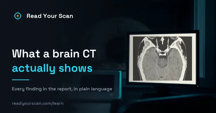
A brain CT shows fresh bleeding, the fluid-filled ventricles and cisterns, the skull, and the gross structure of the brain, in about a minute. It is the fastest way to answer the question that changes emergency treatment. It is much less sensitive than MRI to early stroke and small lesions.
A brain CT (also called a head CT) uses X-rays to make quick, detailed cross-sectional images of the head. This page explains what it looks at and what common report terms mean.
What this scan shows
A brain CT is a fast test that shows the brain tissue, the fluid spaces, the skull, and any bleeding, swelling, or major changes. Doctors often order it urgently after a head injury, for sudden severe headache, dizziness, or stroke-like symptoms, because it is quick and widely available. It is especially good at showing bleeding and bone. The report describes what the images show; your doctor interprets it with your symptoms.

Common findings, in plain words
No acute abnormality
A common and reassuring result meaning no urgent problem such as bleeding was seen. It is one of the most frequent findings on a head CT.
Age-related atrophy
The brain appearing slightly smaller with a bit more space around it, which is a normal part of aging. It is often noted as a routine description.
Small vessel / white matter changes
Changes related to small blood vessels, commonly seen with age. Your doctor considers them alongside your overall vascular health.
Sinus changes
Fluid or thickening in the sinuses, often noticed by chance. These are common and frequently unrelated to the reason for the scan.
Old / chronic changes
Signs of a healed prior event, described as old or chronic. They reflect the past rather than something new. Your doctor explains their meaning.
Calcifications
Small deposits of calcium that are commonly seen in certain areas of the brain and are usually a normal, incidental finding.
Terms you might see
- Acute
- Referring to something new or recent. "No acute finding" means nothing urgent or new was seen.
- Hemorrhage
- Bleeding. A head CT is particularly good at showing whether there is bleeding in or around the brain.
- Atrophy
- A slight reduction in brain volume, often a normal part of aging.
- Ventricles
- The normal fluid-filled spaces inside the brain, whose size is often described.
- Contrast
- A dye sometimes given by vein to highlight certain areas. Many head CTs are done without it.
Questions to ask your doctor
- 1.What prompted this CT, and did the images address that concern?
- 2.Does "no acute finding" mean everything is fine for now?
- 3.Are any findings simply age-related, or does anything stand out?
- 4.Would a different test, such as an MRI, add useful detail?
- 5.Do I need any follow-up, and when?
See these findings on your own scan
Upload your scan (de-identified in your browser) and your radiologist’s report, and every finding is mapped onto the images, explained in plain language and scored — free, no card needed. Informational only — not a diagnosis.
FAQ
- My head CT report says "no acute abnormality" — what does that mean?
- It generally means no urgent problem, such as bleeding, was seen on the scan. It is a common and reassuring result. Your doctor interprets it together with your symptoms and may suggest more if needed.
- Is a brain CT as detailed as an MRI?
- CT is fast and excellent at showing bleeding and bone, which is why it is often used in emergencies. MRI shows soft tissue in finer detail. Your doctor chooses based on what needs to be seen.
- Does a brain CT use a lot of radiation?
- A head CT uses X-rays, and the dose is generally considered low for the information it provides. Your doctor orders it when the benefit of a quick, detailed look outweighs that small exposure.
Related
Go deeper
Sources
- Patient-facing procedure descriptions (MRI, CT, X-ray, ultrasound)RadiologyInfo.org — Radiological Society of North America & American College of Radiology
- CT scan — overview, how it is performed and resultsNHS (United Kingdom)
- Computed tomography (CT) scans and cancerNational Cancer Institute
Read Your Scan is informational only — not a medical diagnosis, and not a substitute for a licensed radiologist or your doctor. If you have urgent symptoms, seek care.