Ultrasound · Abdomen
What an abdominal ultrasound shows, and what the report means
Read Your Scan Editorial Team·Last updated
Written from published radiology references and reviewed for plain language. Not written or signed by a physician, and not a diagnosis. How we write these pages →
The short answer
An abdominal ultrasound shows the liver, gallbladder and bile ducts, the kidneys, the spleen, the aorta and parts of the pancreas, using sound waves rather than radiation. It is excellent for gallstones and fluid, and limited by gas in the bowel and by body habitus.
An abdominal ultrasound uses harmless sound waves — no X-rays — to make live images of the organs in your belly. This page explains what it looks at and what common report terms mean.
What this scan shows
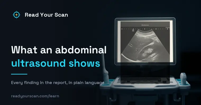
An abdominal ultrasound shows the liver, gallbladder, bile ducts, kidneys, pancreas, spleen, and major blood vessels using sound waves. Doctors often order it for belly pain, to check the gallbladder or liver, to look into abnormal blood tests, or to follow up a known finding. It is safe, painless, and uses no radiation. The report describes what the images show; your doctor interprets it with your symptoms and other tests.

Common findings, in plain words
Gallstones
Stones within the gallbladder, which are common and often cause no symptoms. Your doctor considers whether they relate to how you feel.
Hepatic steatosis (fatty liver)
Extra fat in the liver, seen as the liver appearing brighter than usual. It is common and often improves with lifestyle changes. Your doctor interprets its significance.
Simple renal cyst
A fluid-filled pocket on the kidney. Simple cysts are very common and usually harmless. Your doctor decides whether any check is needed.
Gallbladder wall thickening
The gallbladder wall appearing thicker than usual, which can occur for several reasons. Your doctor considers it together with your symptoms.
Biliary duct dilation
A bile duct appearing wider than usual. It is described so your doctor can decide whether anything further is helpful.
Hemangioma
A common, benign cluster of blood vessels in the liver. These are usually harmless and often just noted for completeness.
Uterine fibroid
A benign growth of the muscular wall of the uterus, extremely common in women of reproductive age. The report gives size and position, which is what determines whether it matters.
Terms you might see
- Echogenicity
- How bright or dark a tissue appears on ultrasound as it reflects sound waves. Doctors compare it to normal tissue.
- Hyperechoic / hypoechoic
- Words for tissue that appears brighter (hyperechoic) or darker (hypoechoic) than usual on the scan.
- Cyst
- A fluid-filled pocket. On ultrasound, simple cysts have a typical, reassuring appearance.
- Bile ducts
- The small tubes that carry bile from the liver and gallbladder toward the intestine.
- Doppler
- An ultrasound mode that shows blood flow, sometimes used to check vessels or organs.
Questions to ask your doctor
- 1.What was the reason for this ultrasound, and did it answer that question?
- 2.If gallstones were seen, do they need treatment or just watching?
- 3.Is a finding like fatty liver something I can improve?
- 4.Do any findings need a follow-up scan or a different test?
- 5.Do the results change my current care in any way?
See these findings on your own scan
Upload your scan (de-identified in your browser) and your radiologist’s report, and every finding is mapped onto the images, explained in plain language and scored — free, no card needed. Informational only — not a diagnosis.
FAQ
- I have gallstones but no pain — do I need them removed?
- Many people have gallstones without any symptoms, and these often do not require treatment. Your doctor considers whether the stones relate to how you feel before discussing any next steps.
- Is an ultrasound safe?
- Yes. Ultrasound uses sound waves rather than X-rays, so there is no radiation. It is painless and widely used, including during pregnancy, which is one reason it is often a first-choice test.
- What does a bright liver on ultrasound mean?
- A liver that appears brighter than usual often reflects extra fat, called fatty liver. It is common and frequently improves with changes to diet and activity. Your doctor interprets what it means for you.
Related
Go deeper
Sources
- Patient-facing procedure descriptions (MRI, CT, X-ray, ultrasound)RadiologyInfo.org — Radiological Society of North America & American College of Radiology
- Medical imaging and radiation dose information for patientsU.S. Food & Drug Administration
Read Your Scan is informational only — not a medical diagnosis, and not a substitute for a licensed radiologist or your doctor. If you have urgent symptoms, seek care.