X-ray interpreter · radiographs, films and phone photos
An AI X-ray interpreter that reads your radiograph — not a chatbot guessing at a photo
Upload a photo of your film or the DICOM file and your X-ray is read in full, straight away, at no cost. You see the study described, the technique named and one line for every finding written about your own image — proof the reading happened. The finished report costs $19 to open.
Your X-ray is read for free · $19 to open the finished report · minutes, not weeks
- A phone photo of the film is enough — no hospital CD required
- Name, IDs and dates stripped in your browser, before upload
- Unreadable photo? Refused, not sold — and it costs you nothing
- Your doctor opens the shared link without an account
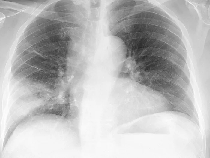
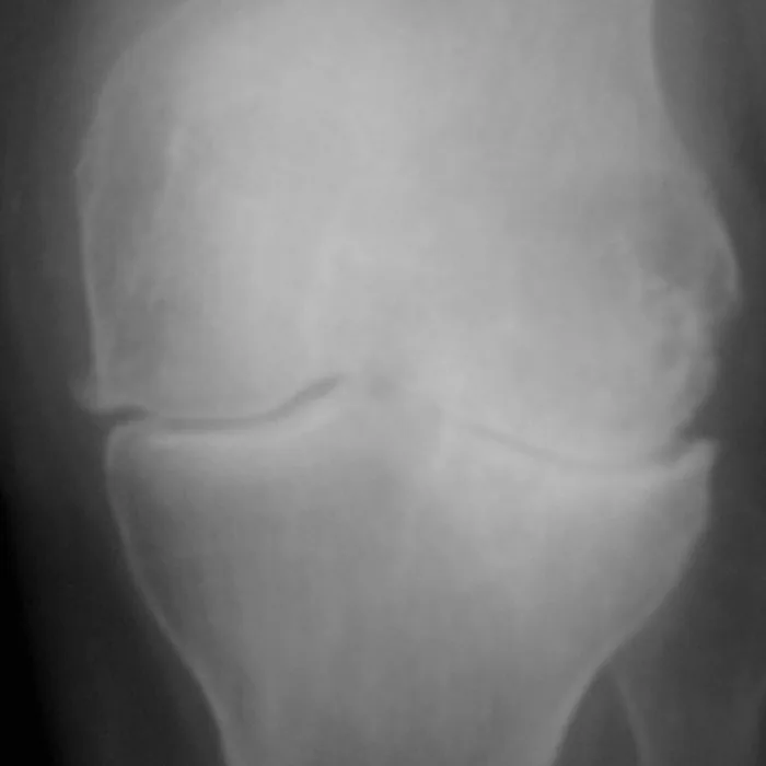
X-ray · chestNeeds attention
“Patchy opacification of the right middle lobe, silhouetting the adjacent right heart border.”
→ A hazy patch low in the right lung, dense enough that the edge of the heart beside it stops showing. What caused it is your doctor’s call.
What happens when you paste an X-ray into ChatGPT
It is the honest comparison, because it is the thing most people try first. A general chatbot will say something about your radiograph. Here is what it is actually doing while it does that — and what a purpose-built X-ray interpreter does instead.
What it opens
A compressed screenshot. It cannot open a DICOM file at all, so the grayscale window your radiologist actually reads on is gone before the model sees anything.
The DICOM if you have one, at its own bit depth and window — or your photo, quality-checked first. What the model reads is the radiograph, not a re-encoded copy of it.
Which side is which
Guessed from the picture. A PA chest film mirrors the patient, and a chatbot reading left-to-right gets the side wrong often enough to matter.
Read from the file’s own metadata and projection, not inferred from the image. Right stays right.
The model behind it
Whichever one the vendor is serving today. No medical-imaging model is reachable from a consumer chat window.
A measured roster — including Google’s MedGemma, gated and unavailable in any chatbot. The reader that wins on flat radiographs is the reader you get.
What it knows about you
Only what you remembered to type into the box.
Your age, sex and the symptoms you described, carried into the reading — plus the radiologist’s report, when you add it.
What you walk away with
A paragraph in a chat window, hedged, unsourced, gone when the thread scrolls. Nothing your doctor can open.
A written report: every finding in plain language with a severity score, placed on your image, plus the specific questions worth asking — on a private link you can send to your doctor.
What it refuses
Very little. A blurry, glared photo gets an answer anyway.
A photo too poor to read is rejected before you are asked for money, and it does not consume anything.
None of this makes the reading a diagnosis. It makes it a reading you can check, share and take to an appointment — which a chat message is not.
The readers
Models a chatbot cannot reach, chosen by measurement
A chatbot hands you whatever single model it happens to be running this month, and you have no say in it. We keep several readers and let measurement decide between them: they are compared head-to-head on real studies with a known diagnosis, and the one that wins on your kind of study is the one that reads yours. That is why a flat radiograph and a volumetric MRI do not go to the same model. The roster includes models built specifically for medical imaging that no chatbot can reach — Google’s MedGemma among them, a gated model you cannot get to from any consumer chat window.
- Claude Opus
- Gemini
- MedGemma 1.5
We do not claim a medical model reads every scan — we claim the winner does, and that we measured which one that is. When a general frontier model beats the medical one on a study type, the general one gets the work.
How reading your X-ray works
A radiograph is a handful of images, so the whole thing can be read before you are asked for anything. That is deliberate: you get to see the work before you decide it was worth paying for.
Step 1 · free
Upload the film — a phone photo will do
Lay the film on a lightbox or against a bright window, fill the frame, avoid glare. JPG, PNG or the DICOM file. Identifying details are stripped in your browser before anything leaves your device.
Step 2 · free
Your X-ray is read in full
The whole study goes to the frontier reader that wins on flat radiographs. Nothing is sampled and nothing is skipped — a chest film is not big enough to need shortcuts.
Step 3 · free
See that it was read
The projection and the anatomy in the field of view are named, and every finding shows its opening line — as many lines as there are findings about your own image. What is withheld is the clinical half of each sentence, not the fact that a sentence exists.
Step 4 · $19
Open the finished report
One credit reveals the report already written for your exam: each finding in full, in plain language, with a severity score, placed on the image, plus follow-up questions grounded in what was found. Ask it questions afterwards at no extra cost.
What the report actually contains
Written for you to read, and built for your doctor to open.
Every finding in plain language
The radiologist’s sentence and what it means, side by side. No “consolidation” without an explanation of what a consolidation is on your film.
A severity score per finding
So routine age-related wear reads differently from the thing that is worth a phone call, instead of everything arriving at the same alarming volume.
The finding placed on your image
Click a finding and the viewer moves to the region it describes. You see the area being talked about, not just its name.
Your report, sentence by sentence
Add the radiologist’s report and every line in it is matched to what is visible on the image and rewritten in plain words.
The questions worth asking
Specific to your findings, not a generic list — the point is to make the next ten minutes with your doctor count.
A private link to share
Send the images and the reading to a doctor or a family member. No account needed on their side, revocable whenever you want.
When an AI X-ray interpreter is worth it
Not always. It earns its place in a few specific situations, and it is fair to say which.
You were handed a film and two dense sentences
The classic X-ray experience: a printed radiograph, a couple of lines of terminology underneath, and no time to ask what any of it meant before the next appointment.
The follow-up is weeks away
You are going to read those sentences a hundred times before anyone explains them. Understanding them now is not a substitute for the appointment — it is what makes the appointment useful.
The report is in a language you do not read
Imaging done abroad, or in a country whose reports come back in a language you never learned. The explanation comes back in yours.
You want a second reading of the images themselves
The reading is done from the images and is deliberately blind to your radiologist’s report. Where the two agree, you can stop worrying. Where they differ is what to ask about.
What this cannot do
A plain radiograph carries far less information than a CT or an MRI. Any tool that pretends otherwise is selling you something.
It is not a diagnosis
Nobody licensed signs it. It explains what is visible in your image; naming a disease, ordering the next test and treating you are your doctor’s job and only your doctor’s.
An X-ray hides more than it shows
Soft tissue, cartilage, discs, early bone injury — a radiograph is largely blind to all of it. The reading says what it cannot see rather than filling the gap with a guess.
A bad photo is a bad reading
Glare, a phone flash on glossy film, half the frame cut off. We refuse photos we cannot read rather than sell you a confident answer built on one.
It has no history but the one you type
Your radiologist has your chart, your previous films and the referral. The reading has the image, your age and sex, and whatever symptoms you chose to describe.
What it costs
One exam
$19
Your X-ray read in full and the finished report opened. One-off payment, no subscription, no account to cancel.
Three examssave 21%
$45
$15 an exam. For a series of follow-up films, or for reading a relative’s scans too. Credits never expire.
- The 3D viewer and the DICOM tools are free, always, with no account
- Reading your radiologist’s report and mapping it to the images is free
- A refused, unreadable photo costs nothing
- Follow-up questions on a report you have opened cost nothing
X-ray questions
- What is an AI X-ray interpreter?
- A tool that reads a radiograph and turns what is visible on it into plain language. Ours describes the study, lists each finding with a severity score, places every finding on the image and ends with the questions worth asking your doctor. It is informational — it interprets the picture for you, it does not diagnose you.
- Why not just paste it into ChatGPT? That is free.
- You can, and many people do. What you get is a general model looking at a compressed screenshot, guessing which side is which, with no access to any medical-imaging model, no report you can hand to a doctor and no record of what was said. Here the file itself is read, the projection and side come from the metadata, the reader is chosen by head-to-head measurement on real studies, and the output is a shareable report. Your X-ray is read here for free too — you pay to open the report, not to be read.
- Can I just photograph my X-ray?
- Yes, and it is the normal way people use this page. Lay the film on a lightbox or hold it against a bright window, fill the frame and avoid glare. A DICOM file gives a better reading when you have one, but a clean photo is enough. If the photo is too poor to read, it is refused before you pay.
- Which model reads it?
- Whichever one wins on flat radiographs in our own head-to-head testing against studies with a known diagnosis — the roster includes Claude Opus, Gemini and Google’s MedGemma, the last of which is a gated medical-imaging model no chatbot can reach. The routing is by study type, which is why an X-ray and an MRI are not sent to the same reader.
- What do I actually see before paying?
- The study and the technique — the projection and what anatomy is in the field of view — plus the opening line of every finding. Six lines under Findings means six findings were written about your image. The clinical half of each sentence is withheld. We do not show you a sample of the reading; we show you that the reading exists.
- Is an AI reading of an X-ray reliable?
- It is informational, not diagnostic. A plain radiograph carries far less information than a CT or MRI, so the reading stays deliberately cautious and tells you what it cannot see. It is not a substitute for a radiologist, and it is at its most useful when it is helping you understand a report a radiologist has already written.
- What happens to my images?
- Identifying details are stripped from the file in your browser, before upload. The images are yours, stored against your account, deletable at any time, and shared only through links you create and can revoke.
Your X-ray gets read either way. The only question is whether you open the report.
Upload the film, watch it be read, and decide afterwards. Nothing is charged until there is a finished report with your name on the study waiting behind the button.
$19 to open the report · not a diagnosis · no subscription
Related
Read Your Scan provides informational explanations only — not a medical diagnosis, and not a substitute for a licensed radiologist or your doctor.