Free online DICOM viewer
Drag in a DICOM folder and view it instantly in 2D and 3D — right in your browser. Nothing is uploaded: the files are decoded locally, so your images stay private on your device. Axial, coronal and sagittal views are MPR reformats rebuilt from the volume, and a slab MIP or MinIP is one click away.
Drop your scan or report here
DICOM, images (JPG/PNG) or a report PDF — add it all together
- Encrypted in transit & at rest
- You control deletion
- We never sell your data
- Results in minutes, not days
Opens: A folder of DICOM (.dcm) files — from a CD, a USB stick or an export. Unzip a zipped export first, then pick the folder. · Files are processed in your browser — nothing is uploaded.
What this tool does
Opens a whole DICOM folder, not one file at a time
Point it at the study folder — or straight at the CD/DVD drive — and it walks every subfolder for you. Hospital exports rarely use tidy .dcm names: files arrive extensionless, UID-named (1.2.840.113619.2…), or with vendor extensions like .ima and .img. All of them load, because the decoder reads file content rather than filenames.
DICOMDIR discs: pick the folder, not the index
The standard hospital disc holds a DICOMDIR index file next to a DICOM folder. Choose the disc, or the folder that contains them, and the images inside are decoded — DICOMDIR itself is only an index, so it is skipped rather than mistaken for a picture.
Every series in the study, listed and switchable
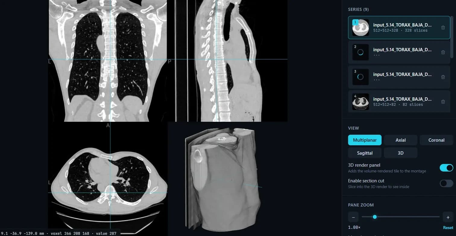
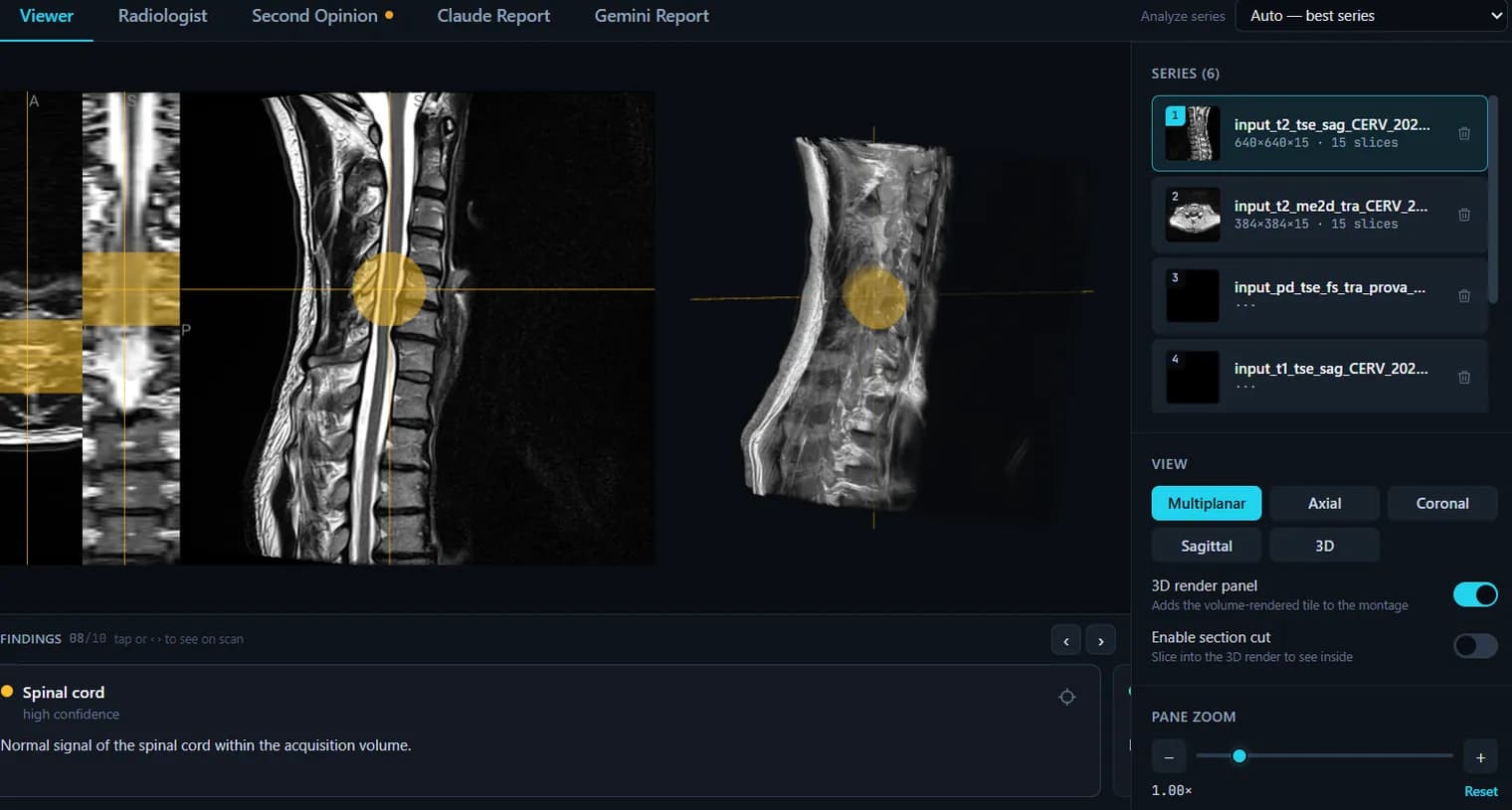
A study is not one image stack. Each series appears in the sidebar with its description and slice count, so you can jump between the sequences, drop the ones you do not need, and keep the scout and localiser images out of the way.
Genuine MPR — axial, coronal and sagittal from the volume
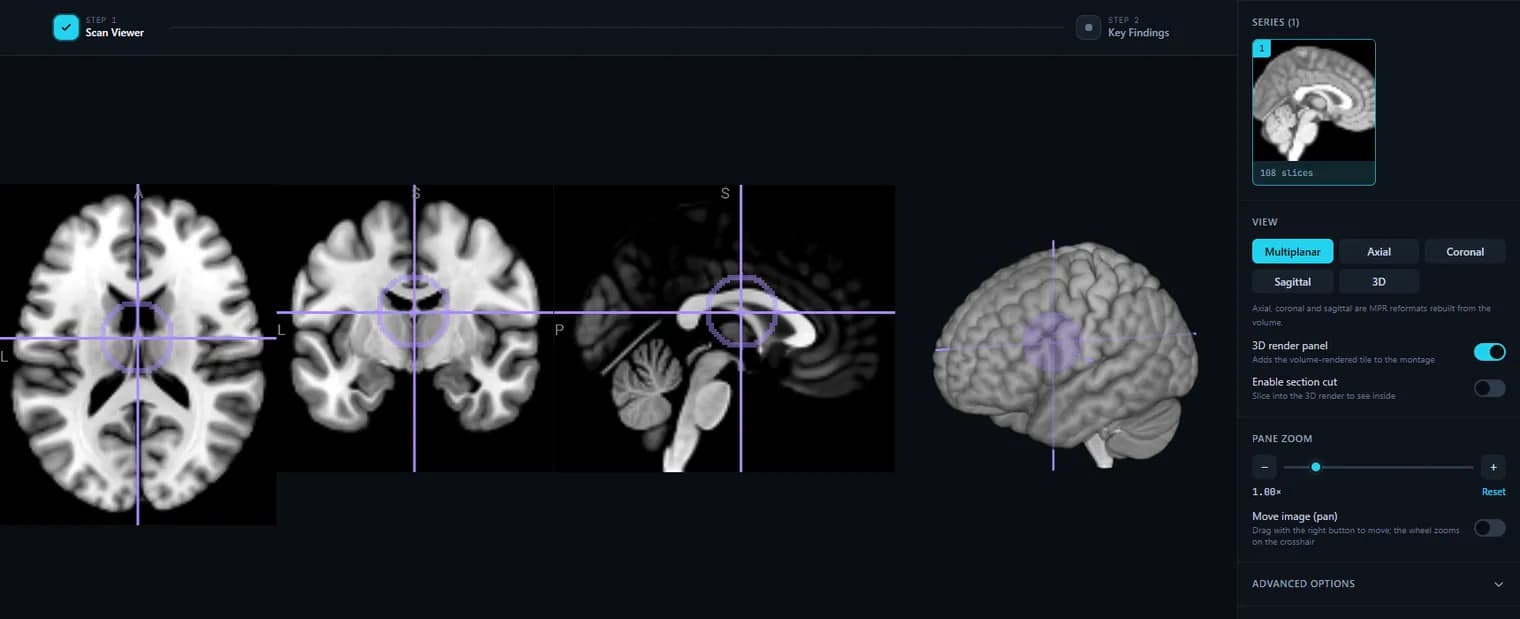
The viewer rebuilds a 3D volume from the slices instead of flipping through pictures, so the coronal and sagittal panes are real multiplanar reformats even when the scan was acquired axially. One crosshair links all three planes: click a point in one and the other two follow.
3D volume rendering, with a section cut
Add the render tile to the layout and rotate it by azimuth and elevation, or set the render depth. Switch on the section cut to clip the volume open and look inside it rather than at its surface.
Slab projections: MIP and MinIP, thickness in millimetres
Under Advanced options you can project a slab instead of a single slice. MIP keeps the brightest voxel through the slab — vessels, contrast, bone; MinIP keeps the darkest — air and airways. You set the thickness in mm, and the projection applies to one plane at a time.
Window and level the way a radiologist does it
Drag on the image with the mouse to set the contrast live, or type an exact min/max intensity range. CT studies get one-click Hounsfield presets (soft tissue, lung, bone, brain, abdomen), and six colormaps — grayscale, bone, hot, inferno, plasma, viridis — are there when grey is not enough.
Reads out millimetres, voxel index and pixel value
A small heads-up display follows the crosshair with the world coordinate in mm, the voxel index, and the raw intensity under it. On a CT that value is the Hounsfield number, which is what tells fat from fluid from calcification.
Navigation built for a laptop as well as a mouse
The wheel scrolls slices, the arrow keys step one slice at a time (hold Shift for ten), the scrubber bar jumps anywhere in the stack, and touch works on a tablet. Right-drag pans in pan mode, the wheel then zooms on the crosshair, and one button resets the view.
Save what you are looking at
Download the current view as a PNG to send to someone, or export the whole volume as a de-identified NIfTI (.nii.gz) — the DICOM identity tags are dropped by the conversion, so the exported file carries the pixels and not the patient.
Runs in the browser, on any computer, with no account
DICOM decoding is dcm2niix compiled to WebAssembly and rendering is WebGL2, so it works in Chrome, Edge, Safari and Firefox on Windows, macOS, Linux and ChromeOS. Nothing to install, nothing to sign up for, and no file is uploaded: your images are decoded on your own machine.
What it does not do
No measurements, ROIs or annotations; no oblique or curved (CPR) reformats; no raw DICOM tag tree — if you need to see the identifying tags, the DICOM anonymizer lists the ones it finds and clears. It is a viewer for understanding your own scan, not a diagnostic-grade reporting workstation. A few compressed transfer syntaxes (JPEG 2000, most often) cannot be decoded in a browser; you get a message saying so and pointing at desktop dcm2niix, not a blank screen.
How to use it
- 1.Click “Upload DICOM folder” and pick the folder from your scan CD or export.
- 2.The series are decoded in your browser (via dcm2niix compiled to WebAssembly).
- 3.Scroll slices, switch planes, rotate the 3D view, and adjust the window.
- 4.Optional: export a de-identified NIfTI copy, or get a plain-language AI report.
FAQ
- Is this DICOM viewer really free?
- Yes. Viewing DICOM files here is completely free and needs no account.
- Are my medical images uploaded to a server?
- No. DICOM files are decoded locally in your browser with WebAssembly, so the pixel data never leaves your device.
- What DICOM files does it support?
- Standard DICOM series from MRI, CT and similar modalities. Some vendor-specific compressed transfer syntaxes may need converting to NIfTI first.
- Can it explain what my scan shows?
- The viewer is free. If you want a plain-language explanation, you can create an account and run an AI report on your uploaded, de-identified scan.
- Can I open the CD or DVD from my hospital?
- Yes. Insert the disc, click “DICOM folder / CD” and choose the CD/DVD drive — the images open right here in your browser, on Windows or Mac, with nothing to install. Your files are never uploaded. If the disc stores its images in a compressed format we can’t read yet (for example JPEG 2000), you’ll get a clear message telling you the next step.
- Does it support MPR and MIP?
- Yes. The viewer reads the whole volume rather than a stack of pictures, so the axial, coronal and sagittal views are genuine MPR reformats. Under Advanced options you can switch on a slab projection and set its thickness in millimetres: MIP keeps the brightest voxel through the slab (vessels, bone), MinIP the darkest (air, airways). Projections work on one plane at a time; oblique and curved (CPR) reformats are not supported yet.
Want your scan explained in plain language?
The viewer is free — and so is understanding your radiologist’s report. Create an account, upload your scan (de-identified in your browser) and your report, and every finding is mapped onto the images, explained in plain language and scored — no card needed. An independent AI reading of the images is a paid extra. Informational only — not a medical diagnosis.