MRI analysis · every series, not one image
AI MRI analysis that reads every series — not the one screenshot you pasted
An MRI is not one picture. It is several series of the same anatomy, each tuned to show something different, which is why the report reads like a foreign language and why a chatbot looking at one frame of it is guessing. Upload the study and it is read series by series, then explained in plain words, scored, and placed on the slice it came from.
Free viewer and free report explanation · $19 for the full AI reading · minutes, not weeks
- The whole DICOM study — hospital CD, USB, ZIP or NIfTI
- Name, IDs and dates stripped in your browser, before upload
- T1, T2, STIR, DWI — the series are detected for you
- Your doctor opens the shared link without an account
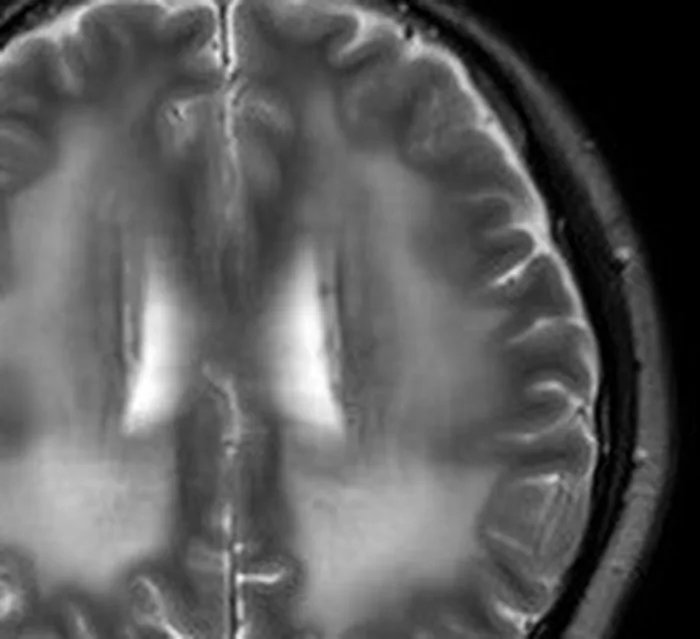
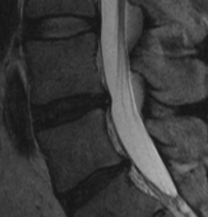
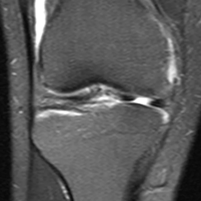
MRI · brainNormal
“Multiple T2-hyperintense foci in the periventricular white matter, of probable microvascular origin.”
→ A few small bright spots in the brain’s wiring, next to the fluid spaces. Very common with age — on their own, not a disease.
What happens when you paste an MRI slice into ChatGPT
This is the free alternative most people try first, so it deserves a straight answer rather than a strawman. A general chatbot will tell you something about that slice. Here is what it is working with while it does.
How much of the exam
The one screenshot you pasted. It cannot open a DICOM folder or scroll a volume, and it has no idea the other series of your exam exist.
The real study, read across every diagnostic series you upload — the T1s, the T2s, the fat-suppressed ones, the diffusion.
Which sequence it is
Guessed from how bright things look. Fluid is dark on T1 and bright on T2, so mistaking one for the other inverts the meaning of what it is describing.
Read from the series metadata, not inferred from contrast. A finding is interpreted against the sequence it actually appears on.
Where it is looking
A flat picture with no coordinates. Which plane, which level and which side are all inferred from what the image looks like.
Plane, slice position and the patient’s left and right come from the scan’s own geometry — the affine, not the appearance.
The model behind it
Whichever one the vendor is serving today. No medical-imaging model is reachable from a consumer chat window.
A measured roster — including Google’s MedGemma, gated and unavailable in any chatbot. The reader that wins on volumetric studies is the reader you get.
What you walk away with
A hedged paragraph in a chat window, gone when the thread scrolls. Nothing your doctor can open.
A written report: every finding in plain language with a severity score, placed on the slice it came from, plus the questions worth asking — on a private link your doctor can open without an account.
Your report
Retyped by you, out of context, if you bother.
Uploaded whole and matched line by line to what is visible in the images. That part is free.
None of this makes the reading a diagnosis. It makes it a reading you can check, share and take to an appointment — which a chat message is not.
The readers
Models a chatbot cannot reach, chosen by measurement
A chatbot hands you whatever single model it happens to be running this month, and you have no say in it. We keep several readers and let measurement decide between them: they are compared head-to-head on real studies with a known diagnosis, and the one that wins on your kind of study is the one that reads yours. That is why a flat radiograph and a volumetric MRI do not go to the same model. The roster includes models built specifically for medical imaging that no chatbot can reach — Google’s MedGemma among them, a gated model you cannot get to from any consumer chat window.
- Claude Opus
- Gemini
- MedGemma 1.5
We do not claim a medical model reads every scan — we claim the winner does, and that we measured which one that is. When a general frontier model beats the medical one on a study type, the general one gets the work.
How an MRI reading works
An MRI is expensive to read properly — many series of real vision tokens — so the credit comes first here and the reading is thorough rather than sampled.
Step 1 · free
Upload the study
The DICOM folder from the hospital CD or USB, a ZIP of it, or a NIfTI file. Drop the whole thing — the series are detected for you. Identifying details are stripped in your browser first.
Step 2 · free
Add the radiologist’s report, if you have one
Every sentence in it is matched to what is visible in your images and rewritten in plain language. This step costs nothing and works on its own, whatever you decide about the reading.
Step 3 · $19
The full AI reading
The study is read series by series, working from the scan’s own geometry, with your age, sex and symptoms in hand. Out comes a report: findings in plain words, each one scored and placed on its slice.
Step 4 · free
Ask it questions, then share it
Follow-up questions on a reading you have paid for cost nothing, and they are grounded in your study rather than in general knowledge. Send the whole thing to your doctor on a private link.
What the reading actually contains
Written for you to read, and built for your doctor to open.
Every series, not a sample
T1, T2, STIR, diffusion — the reading covers the diagnostic series you upload, not one representative image from each.
The vocabulary, decoded
“Hyperintense on T2”, “disc desiccation”, “effusion”. Each term explained where it appears, against your own anatomy rather than in a glossary.
A severity score per finding
So routine age-related wear reads differently from the thing worth a phone call, instead of everything arriving at the same alarming volume.
The finding placed on its slice
Click a finding and the viewer moves to the slice and region it describes. You see the area, not just its name.
Your report, line by line
The radiologist’s sentences matched to what is visible and rewritten in plain words — free, with or without the AI reading.
A private link to share
Send the images and the reading to a doctor or a family member. No account needed on their side, revocable whenever you want.
When an AI MRI reading is worth it
Not always. It earns its place in a few specific situations, and it is fair to say which.
The report is a wall of terminology
The characteristic MRI experience: two paragraphs naming signal intensities and anatomical levels, none of it written for you, and an appointment weeks away.
You cannot tell what is serious
An MRI report of a spine or a knee lists everything visible, including the wear most people your age have. What is missing is which line matters, and that is what the severity score is for.
You want to see where the finding is
“Posterior disc protrusion at L5–S1” means nothing until you are looking at it. Every finding moves the 3D viewer to the region it describes.
You suspect the report missed something
That is a different question, and it has its own page — the second-opinion reading is deliberately blind to your radiologist’s report so the two can be compared.
What this cannot do
The honest half of the pitch. A tool that will not say this is not being straight with you.
It is not a diagnosis
Nobody licensed signs it. It explains what is visible in your study; naming a disease, ordering the next test and treating you are your doctor’s job and only your doctor’s.
It has no prior studies
Half of what a radiologist knows about a finding is how it compares to last year’s scan. The reading sees today’s images only, and says so instead of pretending to a trend.
Findings are not symptoms
Most adults have disc bulges and meniscal wear on an MRI and no pain at all. Whether a finding explains what you feel is a clinical judgement the images alone cannot make.
It has no history but the one you type
Your radiologist has your chart, your examination and the referral. The reading has the images, your age and sex, and whatever symptoms you chose to describe.
What it costs
One exam
$19
One whole MRI study read across its series, with the full report. One-off payment, no subscription, no account to cancel.
Three examssave 21%
$45
$15 an exam. For a follow-up series, or for reading a relative’s studies too. Credits never expire.
- The 3D viewer and the DICOM tools are free, always, with no account
- Reading your radiologist’s report and mapping it to the images is free
- Follow-up questions on a reading you have paid for cost nothing
- Credits never expire and there is nothing recurring to cancel
MRI questions
- What do I upload for an MRI?
- The DICOM folder from the hospital CD or USB stick, a ZIP of it, or a NIfTI file. Drop the whole study — the series are detected for you, and you do not need to work out which one matters.
- Why not just paste a slice into ChatGPT? That is free.
- Because an MRI is not one slice, and it is not one kind of image either. A general chatbot cannot open a DICOM folder, cannot scroll the volume, and has to guess from brightness whether it is looking at a T1 or a T2 — a mistake that inverts the meaning of what it then describes. It also has no access to any medical-imaging model, and produces nothing you can hand to a doctor.
- Which model reads it?
- Whichever one wins on volumetric studies in our own head-to-head testing against studies with a known diagnosis — the roster includes Claude Opus, Gemini and Google’s MedGemma, the last of which is a gated medical-imaging model no chatbot can reach. Routing is by study type: an MRI volume and a flat X-ray do not go to the same reader.
- Is this a diagnosis or a radiologist’s opinion?
- Neither. It is an informational, plain-language explanation of what is visible in your images. Nobody licensed signs it, and it does not replace your radiologist or your doctor.
- How is this different from the second-opinion page?
- This page is about understanding your MRI. The second-opinion reading answers a narrower question — whether an independent read of the same images agrees with your radiologist’s report — and is deliberately blind to that report so the two can be compared line by line. Same engine, different question.
- How much does it cost?
- The viewer is free and explaining your radiologist’s report is free. The full AI reading of the images costs $19 — a credit you buy once, that never expires, and that unlocks one complete reading. Three exams cost $45.
- What happens to my images?
- Identifying details are stripped from the DICOM files in your browser, before upload. The images are yours, stored against your account, deletable at any time, and shared only through links you create and can revoke.
Your MRI was written up for another doctor. Have it written up for you.
Every series read, every finding in plain language, scored and placed where you can see it — and a list of what is actually worth asking at your appointment.
$19 for one exam · explaining your report is free · not a diagnosis
Related
Read Your Scan provides informational explanations only — not a medical diagnosis, and not a substitute for a licensed radiologist or your doctor.