White matter hyperintensities
T2/FLAIR hyperintensities: the bright spots on a brain MRI →The bright spots on your brain MRI, what the report calls them, why "small vessel ischemic disease" sounds worse than it usually is, and how they are graded.Read Your Scan Editorial Team·Last updated
Written from published radiology references and reviewed for plain language. Not written or signed by a physician, and not a diagnosis. How we write these pages →
In plain language
Small bright spots in the brain’s wiring seen on certain images. They are very common, especially with age, and are often related to normal aging or blood-vessel health. Your doctor considers them alongside your symptoms and history.
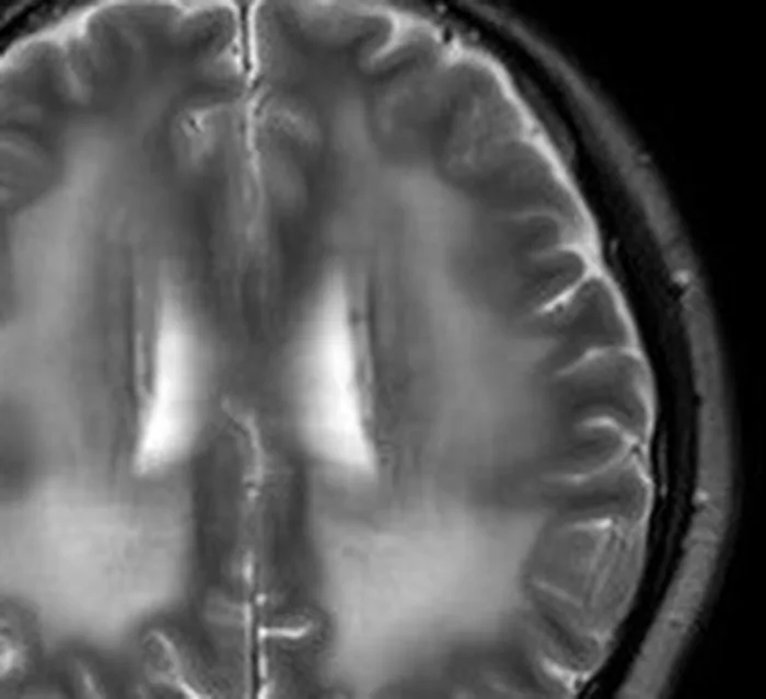
Huang X et al., Front Neurol 2023 · CC BY 4.0
Upload your MRI, CT, X-ray or ultrasound — de-identified in your browser — and every finding is mapped onto your images and explained without jargon.
Where you'll see this
This wording turns up on Brain MRI reports — MRI imaging of the brain. A brain MRI shows the brain tissue in fine detail, along with the fluid spaces, blood vessels, and surrounding structures. Doctors order it to look into headaches, dizziness, memory changes, weakness, seizures, or to follow up on other tests. MRI is especially good at showing soft tissue. Many findings it describes are common, age-related, or minor — the report describes what the pictures show, and your doctor interprets what it means for you.
Questions worth asking your doctor
- What prompted this MRI, and did the images address that concern?
- Are any of the findings expected for my age, or do any stand out?
- Do any findings need a follow-up scan, and if so, when?
- Should these results be discussed with a specialist?
- Is there anything I can do to support my brain and vascular health?
How common is this?
Extremely common with age. White matter hyperintensities (WMH) appear on brain MRI in 50–80 % of people over 65 and in a smaller but measurable proportion of younger adults. They are often labelled “age-related white matter changes” or “small vessel disease”.
When should I worry?
Mild, scattered WMH in an older adult are a normal part of ageing and rarely concerning. They are more significant if extensive for your age, increasing rapidly on serial scans, or accompanied by cognitive symptoms, gait problems or urinary urgency. Many people with significant WMH have no symptoms at all.
What happens next
Mild age-appropriate WMH → reassurance and management of cardiovascular risk factors (blood pressure, diabetes, cholesterol, exercise). Extensive or symptomatic WMH → closer cardiovascular monitoring, possibly cognitive testing. Controlling blood pressure is the single most important modifiable factor.
Frequently asked questions
- Are white matter hyperintensities normal?
- To a degree, yes. Some WMH are a normal part of brain ageing, particularly after 50. They become a concern when more extensive than expected for your age or when accompanied by symptoms like memory problems or difficulty walking.
- Do white matter hyperintensities mean dementia?
- No. While extensive WMH are associated with a higher risk of cognitive decline, most people with WMH do not develop dementia. They are a risk factor, not a diagnosis. Managing cardiovascular health reduces the risk.
- Can white matter hyperintensities be reversed?
- Once present, WMH are generally permanent. However, controlling blood pressure and cardiovascular risk factors can slow or prevent new lesions. Lifestyle interventions (exercise, Mediterranean diet) may slow progression.
- What causes white matter hyperintensities?
- Primarily small vessel disease — reduced blood flow to deep white matter from ageing, hypertension, diabetes and atherosclerosis. In younger patients, migraine and multiple sclerosis are other causes. The pattern helps determine the cause.
Other words on the same report
Where this word comes up
Still worried about what your report says?
Upload your scan (de-identified in your browser) and your radiologist's report, and every finding is mapped onto the images, explained in plain language and scored. Informational only — not a diagnosis.
Read Your Scan is informational only — not a medical diagnosis, and not a substitute for a licensed radiologist or your doctor. If you have urgent symptoms, seek care.