Bone marrow edema (bone bruise)
Read Your Scan Editorial Team·Last updated
Written from published radiology references and reviewed for plain language. Not written or signed by a physician, and not a diagnosis. How we write these pages →
In plain language
A bright area within the bone that often reflects a bruise or stress response. Many resolve over time. Your doctor interprets it with your history.
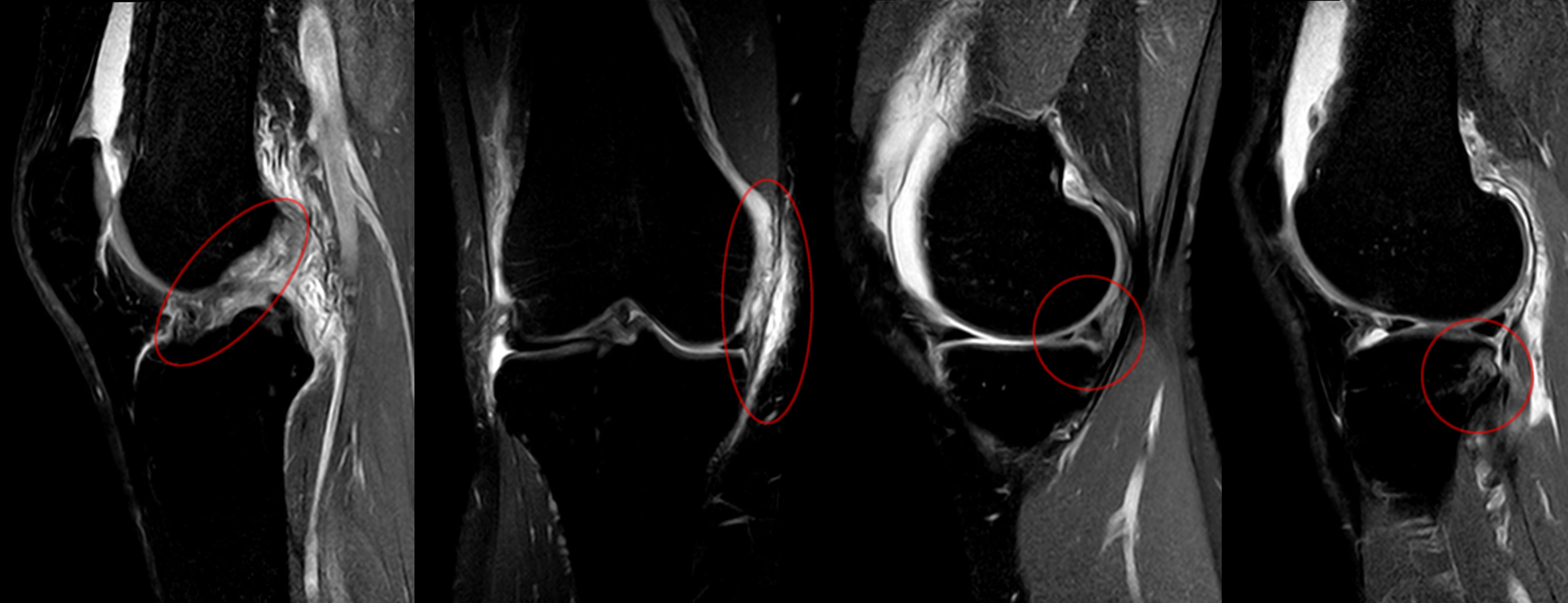
Hellerhoff, Wikimedia Commons · CC BY-SA 3.0
Upload your MRI, CT, X-ray or ultrasound — de-identified in your browser — and every finding is mapped onto your images and explained without jargon.
Where you'll see this
This wording turns up on Knee MRI reports — MRI imaging of the knee. A knee MRI is very good at showing soft tissues that X-rays cannot, including the menisci (the knee’s cushions), the major ligaments, the tendons, the joint cartilage, and the bones. Doctors order it after an injury, for pain, swelling, locking, or instability, or to plan treatment. Some findings, like mild wear, are common with age and may not be the source of symptoms — your doctor connects the images to your exam.
Questions worth asking your doctor
- Do the MRI findings explain the pain, swelling, or instability I feel?
- Is any finding likely age-related rather than from a recent injury?
- What are my treatment options, including non-surgical ones?
- Would physical therapy help before considering other steps?
- Do I need any follow-up, and when should I check back?
How common is this?
Very common on MRI, especially after injury. Bone marrow oedema (bone bruise) is present in 70–80 % of acute knee injuries involving ACL or meniscus tears. In older adults it often accompanies osteoarthritis. It is the MRI signature of stressed or traumatised bone.
When should I worry?
A bone bruise is typically self-limiting and heals over weeks to months. It is concerning if it follows a stress-fracture pattern (suggesting a fracture not visible on X-ray), is extensive with significant cartilage damage, or persists beyond 6–12 months, which may suggest ongoing mechanical stress.
What happens next
Most bone bruises resolve on their own within 2–4 months. Treatment addresses the underlying injury (ACL tear, meniscus tear) rather than the bruise itself. Protected weight-bearing, rest and physical therapy are standard. A follow-up MRI is rarely needed solely for the bone bruise.
Frequently asked questions
- Is a bone bruise the same as a fracture?
- No. A bone bruise means the internal trabecular bone is stressed or has microscopic damage, but the outer cortex is intact. A fracture is a visible break through the cortex.
- How long does a bone bruise take to heal?
- Most resolve in 2–4 months. Larger or more severe bruises can take up to 12 months. During healing you may feel deep, aching pain with weight-bearing.
- Can you walk on a bone bruise?
- Usually yes, though you may need to reduce activity. If the bruise is part of a larger injury, weight-bearing may need to be limited. Your doctor can advise based on location and severity.
- Does a bone bruise show up on X-ray?
- No. Bone bruises are only visible on MRI, which detects changes in marrow fluid. X-rays show the outer cortical bone but cannot see the internal oedema that defines a bone bruise.
Other words on the same report
Where this word comes up
Still worried about what your report says?
Upload your scan (de-identified in your browser) and your radiologist's report, and every finding is mapped onto the images, explained in plain language and scored. Informational only — not a diagnosis.
Read Your Scan is informational only — not a medical diagnosis, and not a substitute for a licensed radiologist or your doctor. If you have urgent symptoms, seek care.