Ground-glass opacity
What “ground-glass opacity” means on a lung CT →A hazy area on a lung CT that does not hide the structures underneath. What causes it, why "ground glass" is a description and not a diagnosis, and what follows.Read Your Scan Editorial Team·Last updated
Written from published radiology references and reviewed for plain language. Not written or signed by a physician, and not a diagnosis. How we write these pages →
In plain language
A hazy area where the lung looks slightly cloudy rather than solid. It can appear with infections, inflammation, or fluid, and often clears up. It is a description of how the tissue looks, not a diagnosis on its own.
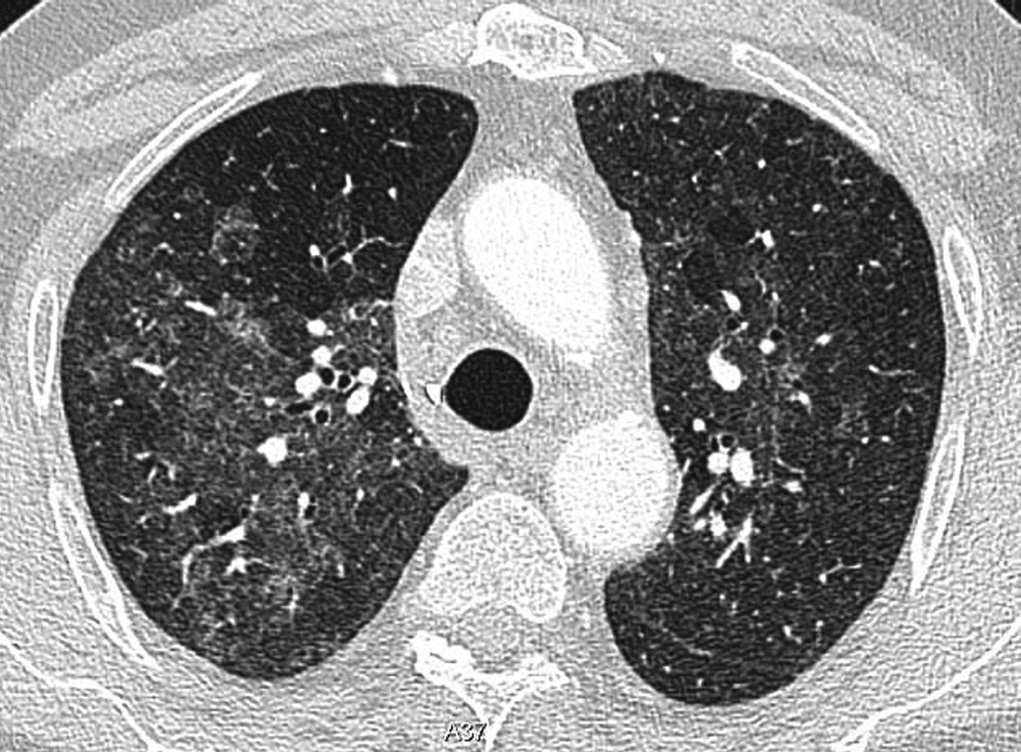
Allen CM et al., Ann Thorac Med 2010 · CC BY 4.0
Upload your MRI, CT, X-ray or ultrasound — de-identified in your browser — and every finding is mapped onto your images and explained without jargon.
Where you'll see this
This wording turns up on Chest CT reports — CT imaging of the chest. A chest CT gives a much more detailed view than a regular chest X-ray. It shows the lungs and airways, the space between the lungs (the mediastinum), lymph nodes, blood vessels, the heart’s outline, and the chest wall. Doctors order it to look more closely at a cough, shortness of breath, an abnormal X-ray, a known nodule, an infection, or to check blood vessels. Seeing something on the images does not by itself mean something is wrong — your doctor reads the pictures together with your history and other tests.
Questions worth asking your doctor
- What was the main reason this chest CT was ordered, and did it answer that question?
- Were any findings unexpected, and do any of them need follow-up?
- If a nodule was seen, does it need another scan later, and when?
- Do the results change anything about my current treatment or medications?
- Is there anything in my history that helps explain what the report describes?
How common is this?
Very common as an incidental finding. Ground-glass opacities (GGOs) appear on chest CTs for many reasons — infection, inflammation, early fibrosis, even breathing artefacts. In lung-cancer screening CTs, small GGOs turn up in up to 20 % of scans. The overwhelming majority are benign.
When should I worry?
A GGO deserves closer attention if it is growing on serial scans, is larger than 20 mm, contains a solid component (part-solid nodule), or is accompanied by persistent cough, weight loss or haemoptysis. A single small pure GGO is usually benign and simply monitored. Words like “stable” and “likely benign” in your report are reassuring.
What happens next
A small pure GGO is typically monitored with a follow-up CT in 6–12 months. If it stays the same, follow-up intervals are extended or stopped. Part-solid or growing GGOs may need further evaluation (PET scan, biopsy, pulmonology referral). Most GGOs turn out to be benign.
Frequently asked questions
- Does ground-glass opacity mean cancer?
- Usually not. GGOs are caused by infections, inflammation, fluid, fibrosis and breathing artefacts, among other things. While some can represent early adenocarcinoma, the vast majority — especially small pure GGOs — are benign.
- What causes ground-glass opacity?
- Common causes include viral or bacterial infections (including post-COVID changes), allergic or inflammatory responses, smoking-related changes, pulmonary oedema and scan artefacts. Only a small fraction represents malignancy.
- How long should a ground-glass opacity be monitored?
- Fleischner Society guidelines recommend follow-up CT at 6–12 months for a suspicious pure GGO. If stable after 5 years, it is generally considered benign and monitoring can stop.
- What is the difference between ground-glass opacity and consolidation?
- A GGO is a hazy area through which underlying lung structures remain visible. Consolidation is denser and completely obscures them. Consolidation usually suggests active infection or inflammation; GGOs are more nonspecific.
Other words on the same report
Where this word comes up
Still worried about what your report says?
Upload your scan (de-identified in your browser) and your radiologist's report, and every finding is mapped onto the images, explained in plain language and scored. Informational only — not a diagnosis.
Read Your Scan is informational only — not a medical diagnosis, and not a substitute for a licensed radiologist or your doctor. If you have urgent symptoms, seek care.