Disc herniation
Read Your Scan Editorial Team·Last updated
Written from published radiology references and reviewed for plain language. Not written or signed by a physician, and not a diagnosis. How we write these pages →
In plain language
A more focal push of disc material beyond its normal border. Many herniations improve on their own over time. The report notes its location so your doctor can relate it to your symptoms.
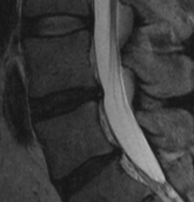
Stillwaterising, Wikimedia Commons · CC0
Upload your MRI, CT, X-ray or ultrasound — de-identified in your browser — and every finding is mapped onto your images and explained without jargon.
Where you'll see this
This wording turns up on Lumbar Spine MRI reports — MRI imaging of the spine. A lumbar (low-back) MRI shows the vertebrae, the cushioning discs between them, the spinal canal, and the nerves that travel to the legs. Doctors order it for low-back pain, sciatica, leg numbness or weakness, or to plan care. Importantly, many of the changes it describes are extremely common and are seen in people with no pain at all — so the report is read together with how you actually feel.
Questions worth asking your doctor
- Do the findings on the MRI match where I actually feel pain or symptoms?
- Are these changes common for my age, or is anything unusual?
- What are my non-surgical options, such as physical therapy?
- Are there any signs that would mean I should seek care sooner?
- Do I need any follow-up imaging, and when?
How common is this?
Common. About 1–3 % of the population has a symptomatic disc herniation at any given time. On MRI, herniated discs appear in roughly 25 % of adults who have no symptoms, so the finding alone does not mean something is wrong.
When should I worry?
Seek urgent attention for sudden loss of bladder or bowel control, numbness in the saddle area (inner thighs and buttocks) or rapidly worsening leg weakness — these suggest cauda equina syndrome, a surgical emergency. Otherwise, a herniation that matches your symptoms is clinically significant but treatable conservatively in most cases.
What happens next
First-line treatment: physical therapy, NSAIDs and activity modification for 6–8 weeks. Most herniations improve on their own — the extruded material can actually shrink as the body reabsorbs it. If pain persists, an epidural steroid injection may be offered. Surgery (microdiscectomy) is effective but usually reserved for persistent or severe cases.
Frequently asked questions
- How long does a disc herniation take to heal?
- Most improve significantly within 6–12 weeks with conservative care. Larger herniations often reabsorb more readily — paradoxically, a bigger herniation can have a better natural course than a small one.
- Is a disc herniation worse than a disc bulge?
- Generally, yes. The inner material has broken through the outer ring, making nerve compression more likely. But size and location matter more than the label — a well-positioned herniation may cause fewer symptoms than a badly-placed bulge.
- Can a herniated disc cause permanent damage?
- Rarely. If nerve compression is severe and left untreated for months, nerve damage can become lasting. This is why persistent weakness or numbness should prompt follow-up rather than waiting.
- Does everyone with a disc herniation need an MRI?
- No. Guidelines recommend MRI only when symptoms persist beyond 6 weeks of conservative care, when red-flag symptoms appear (weakness, bowel or bladder issues), or when surgery is being considered.
Other words on the same report
Where this word comes up
Still worried about what your report says?
Upload your scan (de-identified in your browser) and your radiologist's report, and every finding is mapped onto the images, explained in plain language and scored. Informational only — not a diagnosis.
Read Your Scan is informational only — not a medical diagnosis, and not a substitute for a licensed radiologist or your doctor. If you have urgent symptoms, seek care.