Disc bulge
Read Your Scan Editorial Team·Last updated
Written from published radiology references and reviewed for plain language. Not written or signed by a physician, and not a diagnosis. How we write these pages →
In plain language
A disc that extends a little beyond its normal edge. Bulges are very common and often cause no symptoms. Your doctor considers whether it lines up with what you feel.
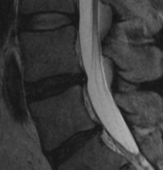
Stillwaterising, Wikimedia Commons · CC0
Upload your MRI, CT, X-ray or ultrasound — de-identified in your browser — and every finding is mapped onto your images and explained without jargon.
Where you'll see this
This wording turns up on Lumbar Spine MRI reports — MRI imaging of the spine. A lumbar (low-back) MRI shows the vertebrae, the cushioning discs between them, the spinal canal, and the nerves that travel to the legs. Doctors order it for low-back pain, sciatica, leg numbness or weakness, or to plan care. Importantly, many of the changes it describes are extremely common and are seen in people with no pain at all — so the report is read together with how you actually feel.
It also appears on Cervical Spine MRI.
Questions worth asking your doctor
- Do the findings on the MRI match where I actually feel pain or symptoms?
- Are these changes common for my age, or is anything unusual?
- What are my non-surgical options, such as physical therapy?
- Are there any signs that would mean I should seek care sooner?
- Do I need any follow-up imaging, and when?
How common is this?
Extremely common. Studies of adults with no back pain find disc bulges in 30–40 % of people over 30 and more than half of those over 50. Most cause no symptoms at all and are discovered incidentally.
When should I worry?
A bulge is significant when it compresses a nerve root and causes matching symptoms — pain, numbness or weakness radiating down a limb. Report phrases that raise the stakes: “nerve root compression”, “foraminal narrowing”, “thecal sac effacement”. A bulge without those is rarely a treatment target.
What happens next
No symptoms → noted and monitored, nothing else. With symptoms the first line is physical therapy and anti-inflammatories for 6–8 weeks. Most bulges improve or become painless within a few months. Surgery is reserved for cases where conservative care fails or neurological deficit is progressing.
Frequently asked questions
- Is a disc bulge the same as a herniated disc?
- No. A bulge means the disc is swelling outward but the outer ring (annulus) is intact. A herniation means the inner material has pushed through the outer ring. Herniations are generally more clinically significant.
- Will a disc bulge heal on its own?
- Often, yes. Many bulges resolve or become asymptomatic within 6–12 months with conservative care. The body can reabsorb some of the protruding material over time.
- Can I exercise with a disc bulge?
- Usually yes, and it is often recommended. Core-strengthening exercises, walking and swimming are typically safe. Avoid heavy lifting and high-impact activities until symptoms improve. Ask your physiotherapist for specific guidance.
- Does a disc bulge mean I need surgery?
- Very rarely. The vast majority are managed without surgery. An operation is considered only when conservative treatment fails after 6+ weeks and neurological symptoms are worsening.
Other words on the same report
Where this word comes up
Still worried about what your report says?
Upload your scan (de-identified in your browser) and your radiologist's report, and every finding is mapped onto the images, explained in plain language and scored. Informational only — not a diagnosis.
Read Your Scan is informational only — not a medical diagnosis, and not a substitute for a licensed radiologist or your doctor. If you have urgent symptoms, seek care.