Free online PET scan viewer
Open a PET or PET-CT study and explore it in 2D and 3D right in your browser. The files are decoded locally — nothing is uploaded — so your images stay private on your device. Axial, coronal and sagittal views are MPR reformats rebuilt from the volume, and a slab MIP or MinIP is one click away.
Drop your scan or report here
DICOM, images (JPG/PNG) or a report PDF — add it all together
- Encrypted in transit & at rest
- You control deletion
- We never sell your data
- Results in minutes, not days
Opens: A folder of DICOM (.dcm) files, or a NIfTI (.nii / .nii.gz) volume. · Files are processed in your browser — nothing is uploaded.
What this tool does
PET and PET-CT studies, series by series
Both the PET and the CT series of a hybrid study load and appear in the list with their descriptions and slice counts, so you can move between the emission images and the anatomical ones.
Colormaps that make uptake readable
Hot, inferno, plasma and viridis turn an intensity range into something the eye separates, which is how PET is normally displayed. Grayscale and bone are there for the CT series.
MIP slabs — the projection PET is usually shown as
A maximum intensity projection through a slab of chosen thickness gives the whole-body look that puts focal uptake into one image instead of across fifty slices. MinIP is available too, and the thickness is set in millimetres.
Multiplanar views and 3D rendering
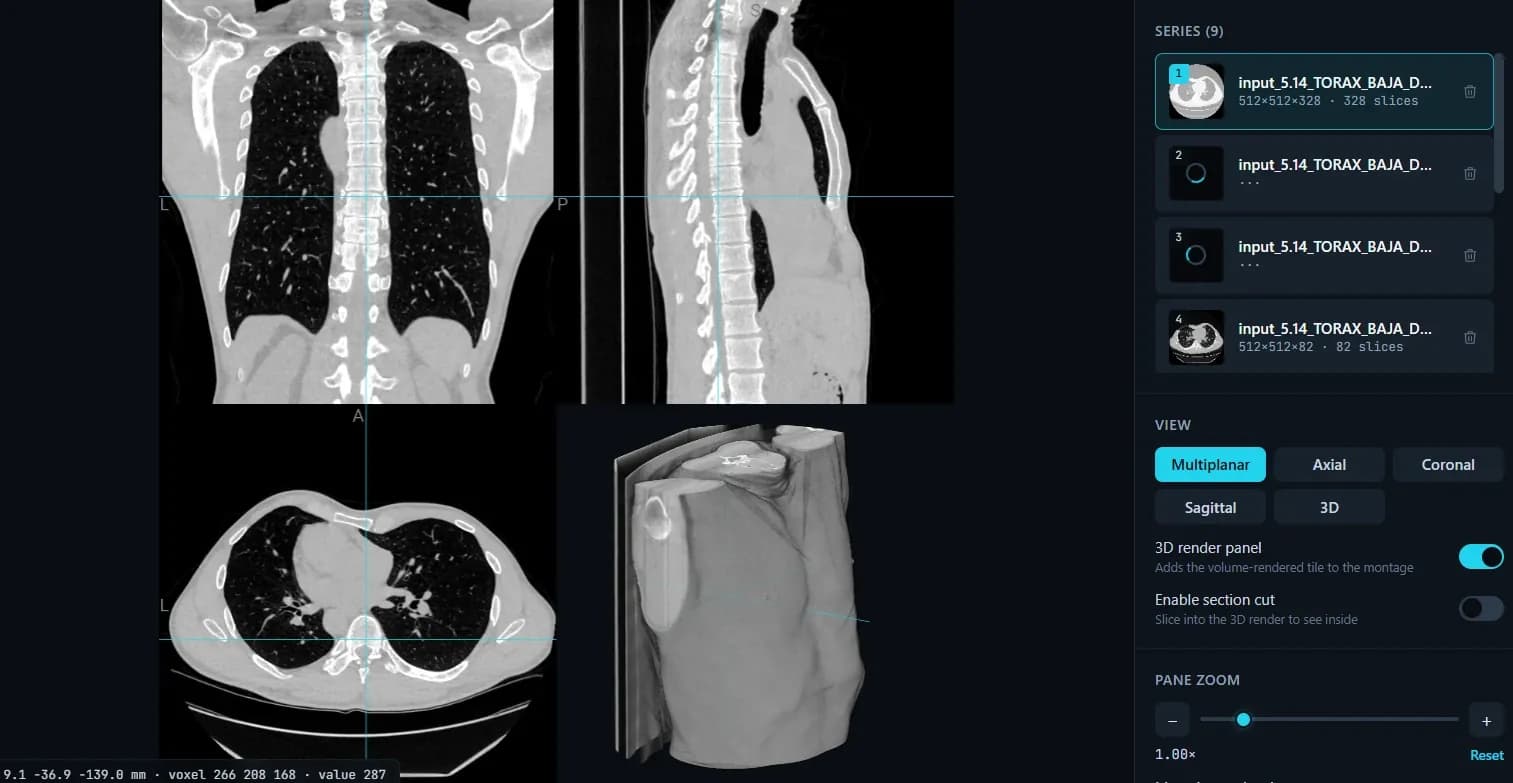
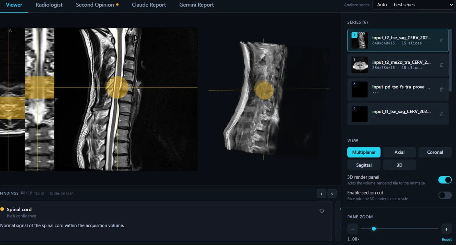
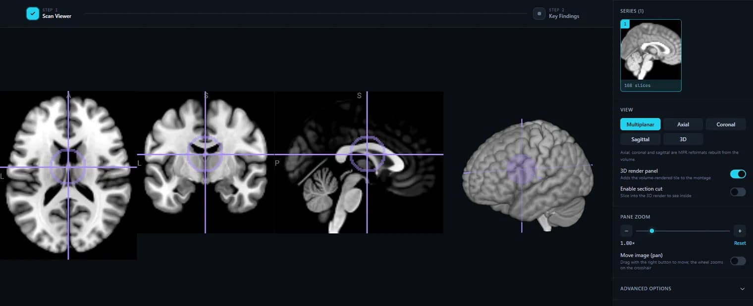
Axial, coronal and sagittal reformats share one crosshair, and a 3D render tile with rotation and a section cut can be added beside them.
Window the intensity range yourself
Set an exact minimum and maximum, or drag on the image. PET display is almost entirely a question of where you clip the upper end of the range, and a fixed default hides either the lesion or the background.
The raw voxel value under the cursor
The readout reports world mm, the voxel index and the stored value at that voxel. Read it as the stored intensity — it is not a computed SUV, and the honest thing to say is that this viewer does not calculate one.
Everything stays on your device
DICOM is decoded in your browser through dcm2niix compiled to WebAssembly; the free viewer uploads nothing.
What it does not do
No SUV computation and no decay or dose correction; no fused PET-on-CT overlay — the two series are viewed one at a time, not blended; no ROI, VOI or lesion measurement. It is a viewer for looking at your own study, not a nuclear medicine reporting workstation.
How to use it
- 1.Click “Upload DICOM folder” and pick your PET (or PET-CT) study.
- 2.It is decoded in your browser and shown in axial, coronal, sagittal and 3D.
- 3.Scroll slices, switch planes, adjust the window, and rotate the 3D view.
FAQ
- Is this PET viewer free?
- Yes. Viewing PET and PET-CT images here is completely free and needs no account.
- Are my PET images uploaded to a server?
- No. The DICOM/NIfTI files are decoded locally in your browser, so the pixel data never leaves your device.
- Can it explain my PET scan?
- The viewer is free. For a plain-language explanation you can create an account and run an AI report on your uploaded, de-identified scan.
- Does it support MPR and MIP?
- Yes. The viewer reads the whole volume rather than a stack of pictures, so the axial, coronal and sagittal views are genuine MPR reformats. Under Advanced options you can switch on a slab projection and set its thickness in millimetres: MIP keeps the brightest voxel through the slab (areas of high uptake), MinIP the darkest (air, airways). Projections work on one plane at a time; oblique and curved (CPR) reformats are not supported yet. It is a slab MIP on the plane you are viewing, not the rotating whole-body MIP a PET workstation shows.
Want your scan explained in plain language?
The viewer is free — and so is understanding your radiologist’s report. Create an account, upload your scan (de-identified in your browser) and your report, and every finding is mapped onto the images, explained in plain language and scored — no card needed. An independent AI reading of the images is a paid extra. Informational only — not a medical diagnosis.